Review
. 2011 May;93(4):275-80.
doi: 10.1308/003588411X571944.

Retroperitoneal tumours: review of management

Affiliations
Review

Retroperitoneal tumours: review of management

Dirk C Strauss et al. Ann R Coll Surg Engl. 2011 May.

Abstract

Introduction: The retroperitoneum can host a wide spectrum of pathologies, including a variety of rare benign tumours and malignant neoplasms that can be either primary or metastatic lesions. Retroperitoneal tumours can cause a diagnostic dilemma and present several therapeutic challenges because of their rarity, relative late presentation and anatomical location, often in close relationship with several vital structures in the retroperitoneal space.

Materials and methods: A comprehensive literature search was conducted using PubMed. Relevant international articles published in the last ten years were assessed. The keywords for search purposes included: retroperitoneum, benign, sarcoma, neoplasm, diagnosis and surgery, radiotherapy, chemotherapy. The search was limited to articles published in English. All articles were read in full by the authors and selected for inclusion based on relevance to this article.

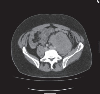
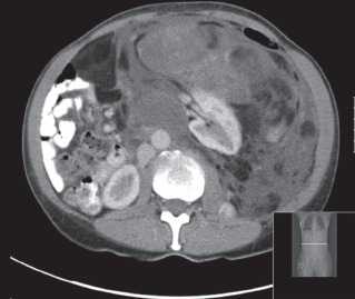
Results: Tumours usually present late and cause symptoms or become palpable once they have reached a significant size. Retroperitoneal tumours are best evaluated with good quality cross-sectional imaging and preoperative histology by core needle biopsy is required when imaging is non-diagnostic. Sarcomas comprise a third of retroperitoneal tumours. Other retroperitoneal neoplasms include lymphomas and epithelial tumours or might represent metastatic disease from known or unknown primary sites. The most common benign pathologies encountered in the retroperitoneum include benign neurogenic tumours, paragangliomas, fibromatosis, renal angiomyolipomas and benign retroperitoneal lipomas.

Conclusions: Complete surgical resection is the only potential curative treatment modality for retroperitoneal sarcomas and is best performed in high-volume centres by a multidisciplinary sarcoma team. The ability completely to resect a retroperitoneal sarcoma and tumour grade remain the most important predictors of local recurrence and disease-specific survival.


Figures

Figure 1
Figure 1
CT of a 35-year-old male showing a large left-sided retroperitoneal dedifferentiated liposarcoma. The tumour weighed 15kg at resection.
Figure 2
Figure 2
Lobulated mass in left iliac fossa that is encasing the distal aorta, bifurcation and left common iliac vessels. A core needle biopsy was consistent with diffuse large B-cell lymphoma.
Figure 3
Figure 3
CT demonstrating a well circumscribed heterogeneous solid mass arising from the left posterior hemipelvis involving the body of the first and second sacral segments from which it is arising. There is scalloping with smooth erosion of the S1 vertebral body on the left, suggesting that this is a longstanding process. A core needle biopsy confirmed this mass to be a benign schwannoma.
Figure 4
Figure 4
Coronal (A) and cross-section (B) CT of a 26-year-old male showing a huge central pelvic mass that arises from the pelvis and extends into the abdomen to the level of the epigastrium. This mass measured 23cm × 19cm × 11cm in diameter. It is displacing bowel loops into the upper abdomen. It is compressing but not obstructing or invading the inferior vena cava and both common iliac and external iliac vessels lie separately from it. The histology was consistent with fibromatosis/desmoid tumour.
Figure 5
Figure 5
CT of a 42-year-old male demonstrating a huge, well differentiated liposarcoma arising from the left retroperitoneum and anteriorly to the left kidney, and extending into the pelvis. The tumour extends superiorly to the left hemidiaphragm, where it passes the midline with displacement of small and large bowel loops into the right flank. The CT attenuation reflects the histological subtype, specifically the amount of fat in the mass, with low-grade, well differentiated liposarcoma entirely or predominantly fatty.
Figure 6
Figure 6
CT of a left dedifferentiated retroperitoneal liposarcoma causing displacement of the left kidney. The CT attenuation reflects the histological subtype with higher grade lesions showing increased density with solid attenuation and contrast enhancement.
Figure 7
Figure 7
CT of an 80-year-old male showing a central abdominal mass probably arising from within the mesentery or a small bowel loop. A core needle biopsy confirmed a gastrointestinal stromal tumour with a mutation found in exon 11 of the KIT gene. The patient was commenced on imatinib.
Figure 8
Figure 8
CT of a 27-year-old male showing a left retroperitoneal mass closely applied to the aorta. A core needle biopsy was consistent with a diagnosis of Ewing's sarcoma and genetic analysis demonstrated a translocation involving the EWSR1 gene. The patient received neoadjuvant chemotherapy and <10% viable tumour was found on post-resection histology.
Figure 9
Figure 9
CT of a radiation-induced right retroperitoneal sarcoma, involving the ribs and diaphragm. Resection included a right nephrectomy, liver wedge resection, resection of the diaphragm and chest wall with mesh reconstruction.

References

    1. Van Roggen JF, Hogendoorn PC. Soft tissue tumours of the retroperitoneum. Sarcoma. 2000;4:17–26. - PMC - PubMed
    1. National Institute for Health and Clinical Excellence. Improving Outcomes for People with Sarcoma: The Manual. London: NICE; 2006.
    1. Weiss SW, Goldblum JR. Enzinger and Weiss's Soft Tissue Tumors. 4th edn. St Louis, Missouri, US: Mosby; 2001.
    1. Strauss DC, Hayes AJ, Thway K, et al. Surgical management of primary retroperitoneal sarcoma. Br J Surg. 2010;97:698–706. - PubMed
    1. Raut CP, Swallow CJ. Are radical compartmental resections for retroperitoneal sarcomas justified? Ann Surg Oncol. 2010;17:1,481–1,484. - PubMed

MeSH terms