. 2011 Dec;104(4):608-19.
doi: 10.1016/j.ymgme.2011.08.018. Epub 2011 Aug 24.

Pathogenesis of aortic dilatation in mucopolysaccharidosis VII mice may involve complement activation

Affiliations

Pathogenesis of aortic dilatation in mucopolysaccharidosis VII mice may involve complement activation

Guilherme Baldo et al. Mol Genet Metab. 2011 Dec.

Abstract

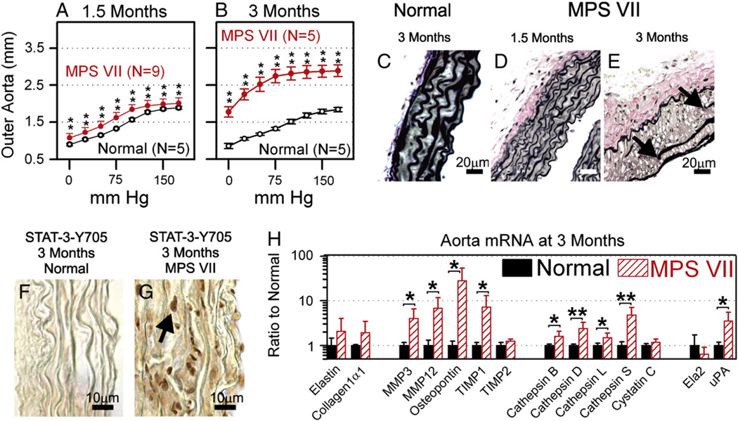
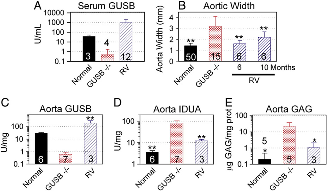
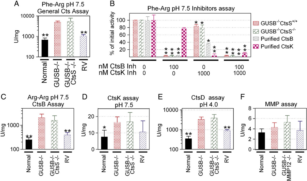
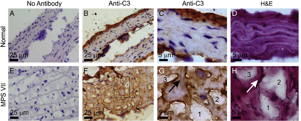
Mucopolysaccharidosis VII (MPS VII) is due to mutations within the gene encoding the lysosomal enzyme β-glucuronidase, and results in the accumulation of glycosaminoglycans. MPS VII causes aortic dilatation and elastin fragmentation, which is associated with upregulation of the elastases cathepsin S (CtsS) and matrix metalloproteinase 12 (MMP12). To test the role of these enzymes, MPS VII mice were crossed with mice deficient in CtsS or MMP12, and the effect upon aortic dilatation was determined. CtsS deficiency did not protect against aortic dilatation in MPS VII mice, but also failed to prevent an upregulation of cathepsin enzyme activity. Further analysis with substrates and inhibitors specific for particular cathepsins suggests that this enzyme activity was due to CtsB, which could contribute to elastin fragmentation. Similarly, MMP12 deficiency and deficiency of both MMP12 and CtsS could not prevent aortic dilatation in MPS VII mice. Microarray and reverse-transcriptase real-time PCR were performed to look for upregulation of other elastases. This demonstrated that mRNA for complement component D was elevated in MPS VII mice, while immunostaining demonstrated high levels of complement component C3 on surfaces within the aortic media. Finally, we demonstrate that neonatal intravenous injection of a retroviral vector encoding β-glucuronidase reduced aortic dilatation. We conclude that neither CtsS nor MMP12 are necessary for elastin fragmentation in MPS VII mouse aorta, and propose that CtsB and/or complement component D may be involved. Complement may be activated by the GAGs that accumulate, and may play a role in signal transduction pathways that upregulate elastases.


Figures

Fig. 1
Fig. 1
Evaluation of MPS VII aortas. A and B. Compliance curves. Aortas were isolated from normal (GUSB+/−, +/+ for other genes) or MPS VII (GUSB−/−, +/+ for other genes) males at 1.5 or 3 months of age, and the average outer diameter of the aorta at the indicated mm Hg ± SD was determined. The Student's t-test was used to compare values in the 2 groups at each pressure, and ** indicates a p valueb0.01. C–E. Elastin stain. Sections of fixed ascending aortas were stained with VVG, which stains elastin a dark color, at the indicated age. The arrows indicate the edges of fragmented elastin fibers in the MPS VII mouse, size bars indicate 20 µm. For all panels, the intima is located on the right side, and the adventitia is on the left side. F and G. Immunostain for tyrosine-phosphorylated STAT3. Frozen sections were stained for the presence of STAT3 that was phosphorylated at tyrosine 705. The arrow identifies the dark nucleus of a positive cell in the MPS VII mouse. Bars indicate 10 µm. H. mRNA levels at 3 months. RNA was isolated from aortas at 3 months of age, and real-time PCR was used to compare levels of genes in normal (N = 6) and MPS VII (N = 5) mice after normalization to β-actin levels. Abbreviations are TIMP for tissue inhibitor of metalloprotease, ELA2 for neutrophil elastase, and uPA for urokinase plasminogen activator.
Fig. 2
Fig. 2
Effect of CtsS and/or MMP12 deficiency on aortic dilatation in MPS VII mice. Aortas were isolated from male mice at 3 months of age, and the outer diameter was determined at the indicated mm Hg of internal pressure. Each line represents a single animal. Black lines represent animals that were never crossed through the CtsS colony, which were derived from either the original MPS VII colony or MPS VII mice that were crossed with MMP12-deficient mice. Red lines represent animals derived from a cross of the MPS VII and the CtsS−/− mice. Blue lines represent animals that were derived from a triple cross of GUSB−/−, CtsS−/−, and MMP12−/− mice. A. Normal. Phenotypically normal mice (GUSB+/− CtsS+/+ MMP12+/+) were analyzed. B. MPS VII mice. GUSB−/− CtsS+/+ MMP12+/+ mice were analyzed C. MPS VII mice with CtsS deficiency. MPS VII mice that were also deficient in CtsS (GUSB−/−, CtsS−/−, MMP12+/+) were analyzed. D. MPS VII mice with MMP12 deficiency. MPS VII mice that also deficient in MMP12 (GUSB−/−, CtsS+/+, MMP12−/−) were analyzed. E. MPS VII mice with CtsS and MMP12 deficiency. MPS VII mice that were also deficient in CtsS and MMP12 (GUSB−/−, CtsS−/−, MMP12−/−) were analyzed. F. Average aortic diameter. The average outer aortic diameter ± SD at 75 mm Hg was determined from the compliance curves in panels A to E. Statistical comparisons were performed between normal mice and other groups; ** indicates a p value<0.01.
Fig. 3
Fig. 3
Effects of gene therapy on aortic abnormalities. Some MPS VII mice were injected IV with 1 × 10¹⁰ transducing units/kg of the RV designated hAAT-cGUSB-WPRE at 2–3 days after birth, while other mice were untreated. A. Serum GUSB activity. GUSB activity was determined at 2 to 6 months of age for normal, untreated MPS VII, and RV-treated MPS VII mice for the indicated number of animals. B. Aortic width. Aortas were dissected and the width was measured at 6 months of age for normal and MPS VII mice, and at 6 months or 10 months as indicated for RV-treated MPS VII mice. C and D. Aorta GUSB and IDUA activity. Extracts prepared at pH 5.5 were tested for GUSB and IDUA activity. E. GAG levels. Extracts prepared at pH 7.5 were tested for GAG levels. Values were compared using ANOVA with Tukey post-hoc analysis, and * represents p ≤ 0.05 and ** represents p ≤ 0.01 as indicated above the bars for comparison with values in untreated MPS VII mice.
Fig. 4
Fig. 4
Cathepsins and MMP assays. Aortas from normal mice, GUSB−/− CtsS+/+ mice (labeled GUSB−/− on the figure), GUSB−/− CtsS−/− mice, or RV-treated MPS VII mice (RV) were homogenized at pH 5.5 for cathepsin assays, while aortas from normal mice, GUSB−/− MMP12+/+ mice (labeled GUSB−/− on figure), GUSB−/− MMP12−/− mice, or RV-treated MPS VII mice (RV) were homogenized at pH 7.5 for the MMP assay. A. Total cathepsin activity at pH 7.5. Samples were incubated with the non-specific substrate Z-Phe-Arg-AMC (Phe-Arg) at pH 7.5, and the activity in U/mg determined. B. Effect of inhibitors on activity against Z-Phe-Arg at pH 7.5. Samples from GUSB−/− CtsS+/+ mice, samples from GUSB−/− CtsS−/− mice with dilated aortas, purified CtsB (0.15 U), or purified CtsK (0.15 U) were incubated with the non-specific Z-Phe-Arg-AMC substrate with and without inhibitors of CtsB and CtsK at the indicated concentration, and the activity relative to that present in samples that did not receive an inhibitor was determined. C. CtsB activity at pH 7.5. Samples were incubated with the CtsB-specific substrate Z-Arg-Arg-AMC (Arg-Arg) at pH 7.5. D. CtsK activity at pH 7.5. Samples were incubated with a CtsK-specific substrate. E. CtsD activity at pH 4.0. Activity for CtsD was determined using a CtsD substrate. F. MMP activity. MMP activity was determined using a commercial kit. For panel B, values in samples that were treated with an inhibitor were compared with values for samples that did not receive the inhibitor using the Student's t test. For other panels, values in other groups were compared with values in untreated MPS VII mice using ANOVA with Tukey post-hoc analysis. * represents p ≤ 0.05 and ** represents p ≤ 0.01.
Fig. 5
Fig. 5
Immunostain for C3. Aortas were from a normal (panels A–D) and an MPS VII (panels E–H) aorta isolated at 6 months. Frozen sections underwent immunostaining without the addition of the anti-C3 first antibody (panels A and E; No Antibody), or with the addition of the anti-C3 first antibody (panels B–C and F–G; Anti-C3) as detailed in the Materials and methods section, and were counterstained with hematoxylin. Panels D and H received hematoxylin and eosin staining only (H&E). The boxes in panels B and F are the regions that are shown at higher power in panels C and G, respectively. Panels G and H represent adjacent sections of the same region of the MPS VII aorta, where 3 separate deposits of GAGs are numbered 1, 2, or 3, and the arrow identifies an elastin fiber. The brown color indicates a positive signal for C3, and nuclei appear blue. Size bars are indicated in each panel. The adventitia is at the top of all low magnification panels.
Fig. 6
Fig. 6
mRNA for complement genes. RNA was extracted from normal (N = 4) and MPS VII (N = 6) mice at 6 months of age and used for reverse transcriptase real-time PCR to assess complement gene mRNA levels. Results are shown as the percentage of β-actin. Genes with expression lower than 0.002% of β-actin were considered not present and are shown as 0.002% in the figure. *p<0.05 or **p<0.01 for comparison of values in MPS VII with those in normal mice using Student's t test. Abbreviations include Fcna for ficolin A, Fcnb for ficolin B, MBL for mannose-binding lectin, MASP for mannan-binding lectin associated serine protease, CD55 for decay accelerating factor, and CD59 for protectin.

References

    1. Neufeld EF, Muenzer J. The mucopolysaccharidoses. In: Scriver CR, Beaudet AL, Sly WS, Valle D, editors. Metabolic and Molecular Basis of Inherited Disease. New York: McGraw Hill; 2001. pp. 3421–3452.
    1. Ausseil J, Desmaris N, Bigou S, Attali R, Corbineau S, Vitry S, Parent M, Cheillan D, Fuller M, Maire I, Vanier MT, Heard JM. Early neurodegeneration progresses independently of microglial activation by heparan sulfate in the brain of mucopolysaccharidosis IIIB mice. PLoS One. 2008;3:1–11. - PMC - PubMed
    1. Simonaro CM, D'Angelo M, He X, Eliyahu E, Shtraizent N, Haskins ME, Schuchman EH. Mechanism of glycosaminoglycan-mediated bone and joint disease: implications for the mucopolysaccharidoses and other connective tissue diseases. Am. J. Pathol. 2008;172:112–122. - PMC - PubMed
    1. Simonaro CM, Ge Y, Eliyahu E, He X, Jepsen KJ, Schuchman EH. Involvement of the Toll-like receptor 4 pathway and use of TNF-alpha antagonists for treatment of the mucopolysaccharidoses. Proc. Natl. Acad. Sci. U. S. A. 2010;107:222–227. - PMC - PubMed
    1. Soliman OI, Timmermans RG, Nemes A, Vletter WB, Wilson JH, ten Cate FJ, Geleijnse ML. Cardiac abnormalities in adults with the attenuated form of mucopolysaccharidosis type I. J. Inherit. Metab. Dis. 2007;30:750–757. - PMC - PubMed

Publication types

MeSH terms