. 2011 Nov;93(5):726-34.
doi: 10.1016/j.exer.2011.09.003. Epub 2011 Sep 17.

FGF signaling regulates rod photoreceptor cell maintenance and regeneration in zebrafish

Affiliations

FGF signaling regulates rod photoreceptor cell maintenance and regeneration in zebrafish

Zhao Qin et al. Exp Eye Res. 2011 Nov.

Abstract

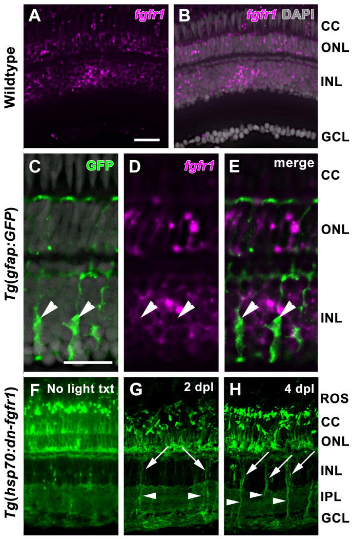
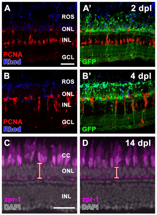
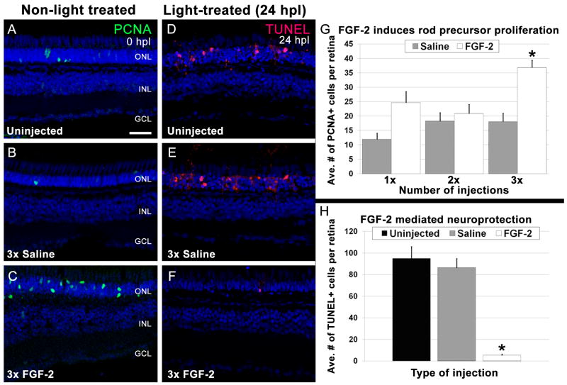
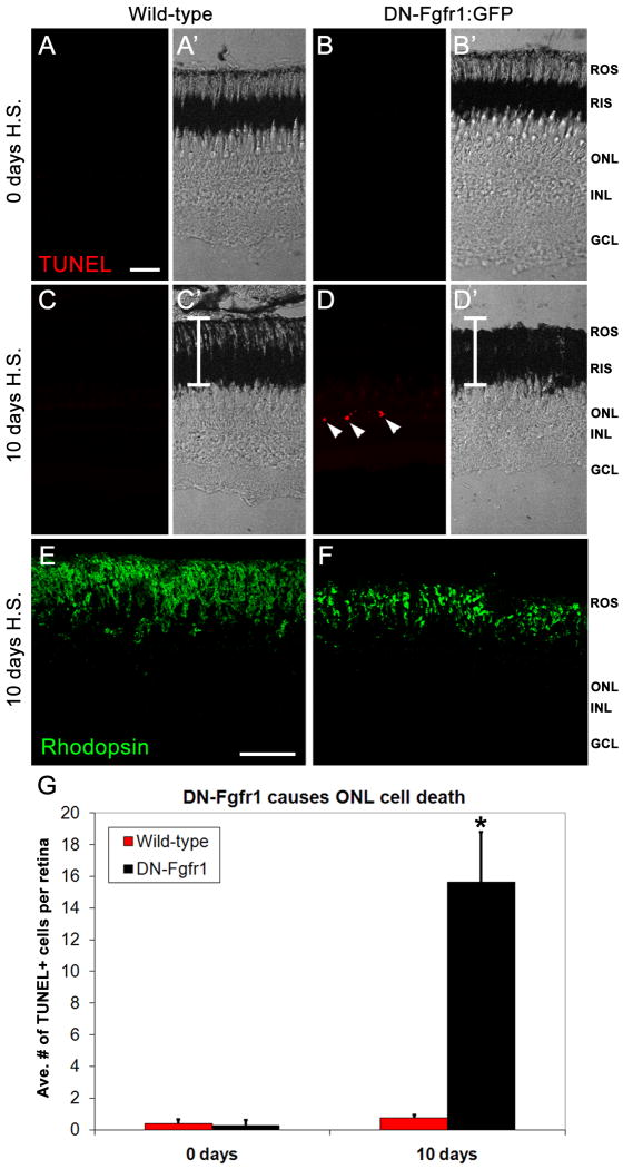
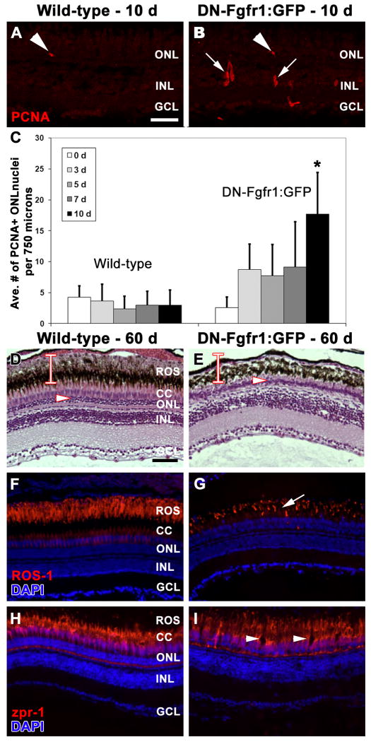
Fgf signaling is required for many biological processes involving the regulation of cell proliferation and maintenance, including embryonic patterning, tissue homeostasis, wound healing, and cancer progression. Although the function of Fgf signaling is suggested in several different regeneration models, including appendage regeneration in amphibians and fin and heart regeneration in zebrafish, it has not yet been studied during zebrafish photoreceptor cell regeneration. Here we demonstrate that intravitreal injections of FGF-2 induced rod precursor cell proliferation and photoreceptor cell neuroprotection during intense light damage. Using the dominant-negative Tg(hsp70:dn-fgfr1) transgenic line, we found that Fgf signaling was required for homeostasis of rod, but not cone, photoreceptors. Even though fgfr1 is expressed in both rod and cone photoreceptors, we found that Fgf signaling differentially affected the regeneration of cone and rod photoreceptors in the light-damaged retina, with the dominant-negative hsp70:dn-fgfr1 transgene significantly repressing rod photoreceptor regeneration without affecting cone photoreceptors. These data suggest that rod photoreceptor homeostasis and regeneration is Fgf-dependent and that rod and cone photoreceptors in adult zebrafish are regulated by different signaling pathways.


Figures

Figure 1
Figure 1. Expression of fgfr1 and hsp70:dn-fgfr1 in the adult zebrafish retina
A. Expression of fgfr1 transcripts in both rod and cone photoreceptors and in the inner nuclear layer is detected by in situ hybridization. B. Overlay of panel A with DAPI nuclear staining. C. GFP expression in Müller glia (arrowheads) in the Tg(gfap:GFP)mi2002 retina. D. Expression of fgfr1 in the same retinal section shown in panel C. E. Expression of fgfr1 in the inner nuclear layer does not colabel with GFP-positive Müller glia (arrowheads). F. Ubiquitous GFP expression driven by the hsp70 promoter in a Tg(hsp70:dn-fgfr1) retina two days after daily heat shock. G. Strong expression of dn-fgfr1-GFP fusion protein in Müller glia soma (arrows) and cell processes (arrowheads) at 2 days of constant light treatment (2 dpl). H. Strong expression of dn-fgfr1-GFP fusion protein in Müller glia soma (arrows) and cell processes (arrowheads) at 4 days of constant light treatment (4 dpl). ROS = rod outer segments; CC = cone cells; ONL = outer nuclear layer; INL = inner nuclear layer; IPL = inner plexiform layer; GCL = ganglion cell layer. Scale bar: Panel A = 50 μm (A-B, F-H); Panel C = 25 μm (C-E).
Figure 2
Figure 2. Fgf signaling is required for rod, but not cone, photoreceptor cell regeneration
A. Retinal section from a Tg(hsp70:dn-fgfr1) adult zebrafish following daily heat shock and 2 days of constant light treatment (2 dpl). Müller glia cell proliferation (PCNA immunolabeling; red) and rod outer segment degeneration (Rhodopsin immunolabeling; blue) are unaffected by loss of Fgf signaling. A'. Overlay of panel A showing immunolocalization of the dn-fgfr1-GFP fusion protein. B. Retinal section from a Tg(hsp70:dn-fgfr1) adult zebrafish following daily heat shock and 4 days of constant light treatment (4 dpl). The continual proliferation of inner nuclear layer progenitors (PCNA immunolabeling; red) and their migration to the outer nuclear layer is unaffected by loss of Fgf signaling. B'. Overlay of panel B showing immunolocalization of the dn-fgfr1-GFP fusion protein. C – D. Retinal sections from wild-type (C) and Tg(hsp70:dn-fgfr1) zebrafish (D) 14 days after intense light damage (14 dpl) and daily heat shock. Double cones are immunolabeled with zpr-1 (magenta). DAPI nuclear stain shows retinal layers. C. Wild-type retinas contain zpr-1+ double cones and thick outer nuclear layer (red bar), which primarily houses rod photoreceptor nuclei. D. The Tg(hsp70:dn-fgfr1) retinas contain zpr-1+ double cones and a thinner than normal outer nuclear layer (red bar). ROS = rod outer segments; CC = cone cells; ONL = outer nuclear layer; INL = inner nuclear layer; GCL = ganglion cell layer. Scale bar: Panel A = 50 μm (A-B'); Panel C = 50 μm (C-D).
Figure 3
Figure 3. FGF-2 induces rod precursor proliferation and provides neuroprotection
A - C. Retinal sections from undamaged eyes that were either uninjected (A), or injected three consecutive days with Saline (B) or FGF-2 (C). PCNA immunolocalization (green) shows proliferating cells. Nuclei are stained with TO-PRO-3 (blue). A. A few PCNA-positive rod precursors are present in the outer nuclear layer. B. Saline injections did not significantly increase the number of PCNA-positive rod precursors. C. Large numbers of PCNA-positive rod precursors are observed in the outer nuclear layer in FGF-2-injected retinas. D - F. Retinal sections of 24-hour light-treated zebrafish that were either uninjected (D) or injected three consecutive days with Saline (E) or FGF-2 (F). TUNEL immunolocalization (red) shows cells undergoing apoptosis. Nuclei are stained with TO-PRO-3 (blue). D. Large numbers of TUNEL-positive cells are observed in the outer nuclear layer of uninjected retinas. E. Saline injections did not significantly alter the number of TUNEL-positive nuclei. F. Eyes injected three consecutive days with FGF-2 exhibited significantly fewer TUNEL-positive cells. G. Graphic representation of the average number of PCNA-positive cells in the outer nuclear layer in non-light treated eyes injected with either Saline or FGF-2. H. Graphic representation of the average number of TUNEL-positive cells in 24-hour light-treated retinas that were either uninjected, or injected for three consecutive days with Saline or FGF-2.
Figure 4
Figure 4. Loss of Fgf signaling causes rod photoreceptor apoptosis and outer segment degeneration
A-A'. Fluorescent and phase contrast images showing the absence of any TUNEL labeling (red) in a wild-type retina prior to heat-shock. B-B'. Fluorescent and phase contrast images showing the absence of TUNEL labeling (red) in a Tg(hsp70:dn-fgfr1) retina prior to heat-shock. C-C'. Fluorescent and phase contrast images showing the absence of TUNEL labeling (red) in a wild-type retina after 10 days of daily heat-shock. The white bar depicts the normal thickness of rod inner and outer segments. D-D'. Fluorescent and phase contrast images showing TUNEL-positive nuclei in the outer nuclear layer (red) of a Tg(hsp70:dn-fgfr1) retina after 10 days of daily heat-shock. The white bar depicts the normal thickness of rod inner and outer segments, showing rod outer segment degeneration in transgenic retinas. E. Rhodopsin immunolocalization to rod outer segments in a wild-type retina after 10 days of daily heat-shock. F. Rhodopsin immunolocalization to rod outer segments in a Tg(hsp70:dn-fgfr1) retina after 10 days of daily heat-shock, showing degenerated and disorganized outer segments. G. Graphic depiction of the average number of TUNEL-positive nuclei in wild-type and Tg(hsp70:dn-fgfr1) retinas after either 0 or 10 days of daily heat shock. ROS = rod outer segments; RIS = rod inner segments; ONL = outer nuclear layer; INL = inner nuclear layer; GCL = ganglion cell layer. Scale bar: Panel A = 50 μm (A-D'); Panel E = 50 μm (E-F).
Figure 5
Figure 5. Long-term defect in Fgf signaling results in significant loss of rod, but not cone, photoreceptors
A. PCNA immunolocalization (red) in a wild-type retina after 10 days of daily heat-shock shows isolated rod precursor cell proliferation in the ONL (arrowhead). B. PCNA immunolocalization (red) in a Tg(hsp70:dn-fgfr1) retina after 10 days of daily heat-shock shows increased proliferation in rod precursor cells (arrowhead; Panel C) and in inner nuclear layer progenitor cells (arrows). C. Graphic depiction comparing the average number of PCNA-positive cells observed in the outer nuclear layer in wild-type and Tg(hsp70:dn-fgfr1) retinas following daily heat-shock over multiple time points. D-I. Wild-type (D, F, H) and Tg(hsp70:dn-fgfr1) (E, G, I) retinas following 60 days of daily heat-shock. D. Histological section of a wild-type retina showing the normal outer nuclear layer (arrowhead) and the thickness of the rod inner and outer segments (bar). E. Histological section of a Tg(hsp70:dn-fgfr1) retina, showing a thin outer nuclear layer (arrowhead) and a near complete loss of rod outer segments (compare the bar in D and E). F. ROS-1 immunolabeling (red) overlaid with DAPI nuclear stain (blue) in a wild-type retina, showing normal rod outer segment thickness and organization. G. A corresponding Tg(hsp70:dn-fgfr1) retinal section, showing ROS-1 immunolocalization (red) to truncated and disorganized rod outer segments (arrow). H. Immunolocalization of zpr-1 to double cones (red) overlaid with DAPI nuclear stain (blue) in a wild-type retina. I. A corresponding Tg(hsp70:dn-fgfr1) retinal section, showing zpr-1 immunolocalization to double cones (red), which are present, but exhibit areas of disorganization (arrowheads). ROS = rod outer segments; CC = cone cells; ONL = outer nuclear layer; INL = inner nuclear layer; GCL = ganglion cell layer. Scale bar: Panel A = 50 μm (A-B'); Panel D = 50 μm (D-I).

Similar articles

Cited by

References

    1. Becker T, Wullimann MF, Becker CG, Bernhardt RR, Schachner M. Axonal regrowth after spinal cord transection in adult zebrafish. J Comp Neurol. 1997;377:577–595. - PubMed
    1. Bernardos RL, Barthel LK, Meyers JR, Raymond PA. Late-stage neuronal progenitors in the retina are radial Muller glia that function as retinal stem cells. J Neurosci. 2007;27:7028–7040. - PMC - PubMed
    1. Bernardos RL, Raymond PA. GFAP transgenic zebrafish. Gene Expr Patterns. 2006;6:1007–1013. - PubMed
    1. Boilly B, Cavanaugh KP, Thomas D, Hondermarck H, Bryant SV, Bradshaw RA. Acidic fibroblast growth factor is present in regenerating limb blastemas of axolotls and binds specifically to blastema tissues. Dev Biol. 1991;145:302–310. - PubMed
    1. Cai Z, Feng GS, Zhang X. Temporal requirement of the protein tyrosine phosphatase Shp2 in establishing the neuronal fate in early retinal development. J Neurosci. 2010;30:4110–4119. - PMC - PubMed

Publication types

MeSH terms

Substances

LinkOut - more resources