. 2011 Oct 11;21(19):1635-40.
doi: 10.1016/j.cub.2011.08.029. Epub 2011 Sep 22.

MRI magnetic field stimulates rotational sensors of the brain

Affiliations

MRI magnetic field stimulates rotational sensors of the brain

Dale C Roberts et al. Curr Biol. .

Abstract

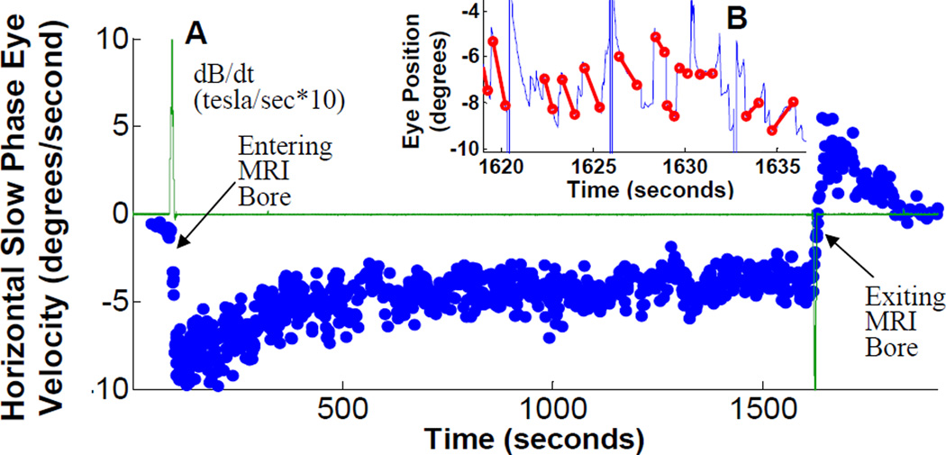
Vertigo in and around magnetic resonance imaging (MRI) machines has been noted for years [1, 2]. Several mechanisms have been suggested to explain these sensations [3, 4], yet without direct, objective measures, the cause is unknown. We found that all of our healthy human subjects developed a robust nystagmus while simply lying in the static magnetic field of an MRI machine. Patients lacking labyrinthine function did not. We use the pattern of eye movements as a measure of vestibular stimulation to show that the stimulation is static (continuous, proportional to static magnetic field strength, requiring neither head movement nor dynamic change in magnetic field strength) and directional (sensitive to magnetic field polarity and head orientation). Our calculations and geometric model suggest that magnetic vestibular stimulation (MVS) derives from a Lorentz force resulting from interaction between the magnetic field and naturally occurring ionic currents in the labyrinthine endolymph fluid. This force pushes on the semicircular canal cupula, leading to nystagmus. We emphasize that the unique, dual role of endolymph in the delivery of both ionic current and fluid pressure, coupled with the cupula's function as a pressure sensor, makes magnetic-field-induced nystagmus and vertigo possible. Such effects could confound functional MRI studies of brain behavior, including resting-state brain activity.


Figures

Figure 1
Figure 1
(A) Slow-phase velocity (SPV) during 25 minute trial. Data from a representative subject showing typical response. Subject is initially outside MRI bore at left of figure. EMI voltage (green line) peaks positive during subject movement into bore, and negative near end of trace as subject moves out of bore. SPV (+right, −left) peaks after in bore, settles to steady state after about 10 minutes, and reverses upon removal from bore. Inset (B) shows several seconds of the original eye position data during bore exit from which the eye velocity is derived. Slow phases are marked with a red line, and the slope of each line becomes a single blue dot on the velocity trace. The change in direction of slopes corresponds to the change in sign of SPV during bore exit. (Supplemental Figure S1 shows head position inside and outside the MRI bore, and data from patients with bilateral vestibular loss; Movie S1 shows typical eye movements during bore entry)
Figure 2
Figure 2
Slow-phase velocity (SPV) is related to static head pitch position (+pitch, chin towards chest). (A) Data from subject S1 in separate trials during a single session, obtained in the order shown from left to right. The first position was repeated at the end of the session to demonstrate the robust repeatability of the phenomenon. (B) SPV data for all ten subjects. Each data point is the average SPV over 45 seconds after the subject is completely in the bore (with standard deviation error bars). Shows consistent relationship between SPV and head pitch angle for all ten subjects (traces labeled for each subject, S1 through S10), yet reveals considerable variation in head pitch angle where SPV null occurs (where each subject line crosses the horizontal zero SPV axis). Range is from −27° for subject S6, to +32° for subject S1.
Figure 3
Figure 3
(A–D) Stimulation is due to a static mechanism. All data plots show SPV (blue dots, deg/sec) and EMI search coil voltage (green trace, tesla/sec*10, except panel A, tesla/sec*5) over time in seconds. Data shows that eye movements are not related to transient, dynamically induced EMI voltage. (E) Stimulation is due to a polarity sensitive mechanism. The SPV direction reverses when the magnetic field vector is reversed relative to the head. (see also Supplemental Data Figure S2)
Figure 4
Figure 4
Geometric model using Lorentz forces. (A) Right-hand rule relationship among current (green), magnetic field (yellow), and resulting Lorentz force (red). (B) Two-dimensional view of lateral canals, ampulla, and utricle, looking through top of head (vertical canals not shown), in head pitch-up position, with resulting Lorentz forces to the left (same orientation as panel C). The sign of the utricular force contribution depends on head pitch in the magnetic field as shown in C and D. (C) Two three-dimensional views of the same head pitch up position (utricle current vector pointing slightly upward), with resulting utricular Lorentz force to subject left. (D) Head pitch down (utricle current vector pointing slightly down), and utricular Lorentz force to subject right. (see also Supplemental Figure S3)

Comment in

References

    1. Schenck JF. Human exposure to 4.0-Tesla magnetic fields in a whole-body scanner. Med. Phys. 1992;19:1089. - PubMed
    1. Heilmaier C, Theysohn JM, Maderwald S, Kraff O, Ladd ME, Ladd SC. A large-scale study on subjective perception of discomfort during 7 and 1.5T MRI examinations. Bioelectromagnetics. 2011 - PubMed
    1. Schenck JF. Health and Physiological Effects of Human Exposure to Whole-Body Four-Tesla Magnetic Fields during MRI. AnnNYAcad. Sci. 1992;649:285–301. - PubMed
    1. Glover PM, Cavin I, Qian W, Bowtell R, Gowland PA. Magnetic-field-induced vertigo: A theoretical and experimental investigation. Bioelectromagnetics. 2007;28:349–361. - PubMed
    1. Marcelli V, Esposito F, Aragri A, Furia T, Riccardi P, Tosetti M, Biagi L, Marciano E, Di Salle F. Spatio-temporal pattern of vestibular information processing after brief caloric stimulation. Eur. J. Radiol. 2009;70:312–316. - PubMed

Publication types