. 2011 Nov;179(5):2443-53.
doi: 10.1016/j.ajpath.2011.07.049. Epub 2011 Sep 21.

LMNA mutations induce a non-inflammatory fibrosis and a brown fat-like dystrophy of enlarged cervical adipose tissue

Affiliations

LMNA mutations induce a non-inflammatory fibrosis and a brown fat-like dystrophy of enlarged cervical adipose tissue

Véronique Béréziat et al. Am J Pathol. 2011 Nov.

Abstract

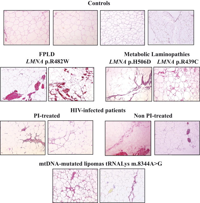
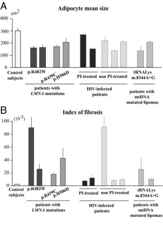
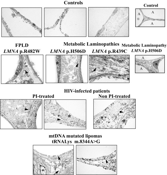
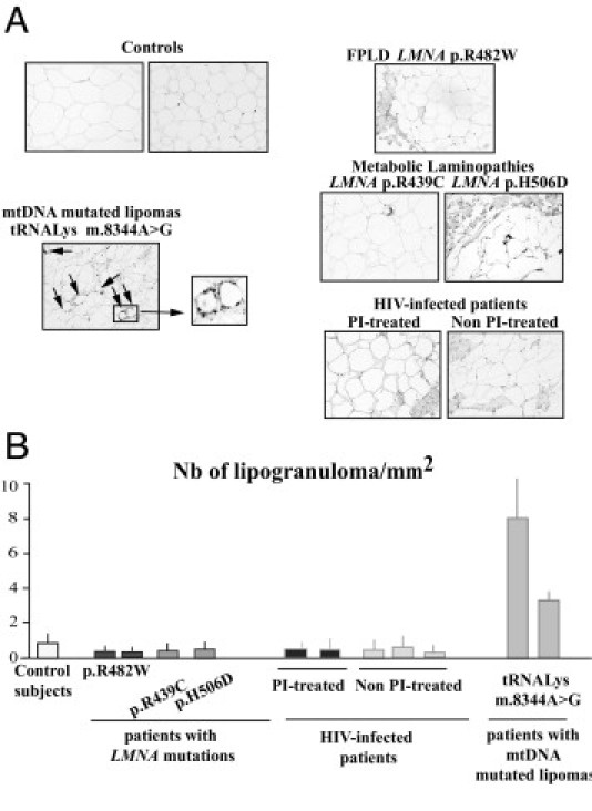
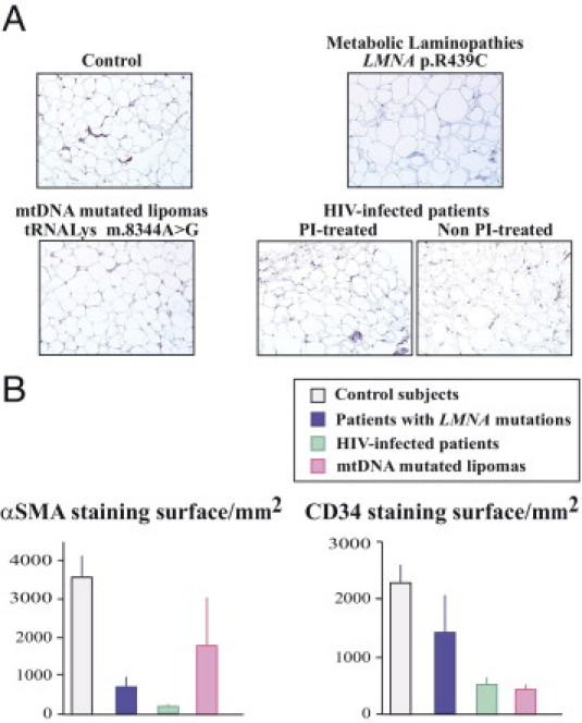
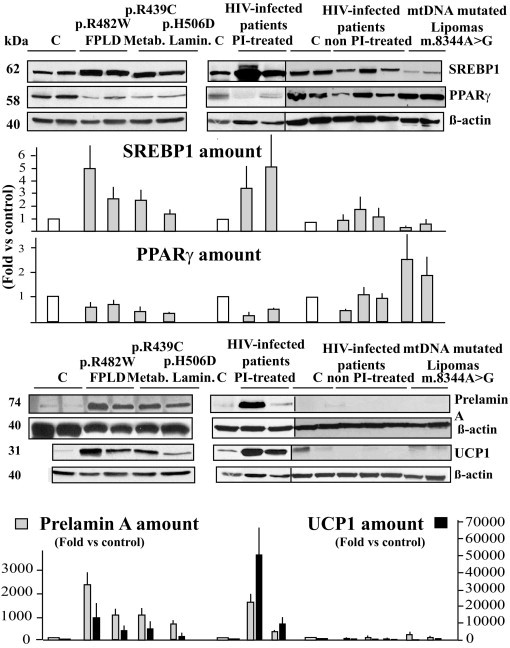
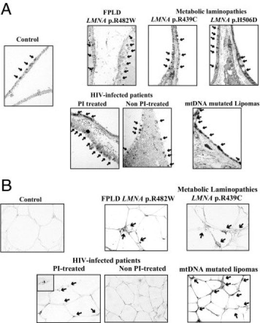
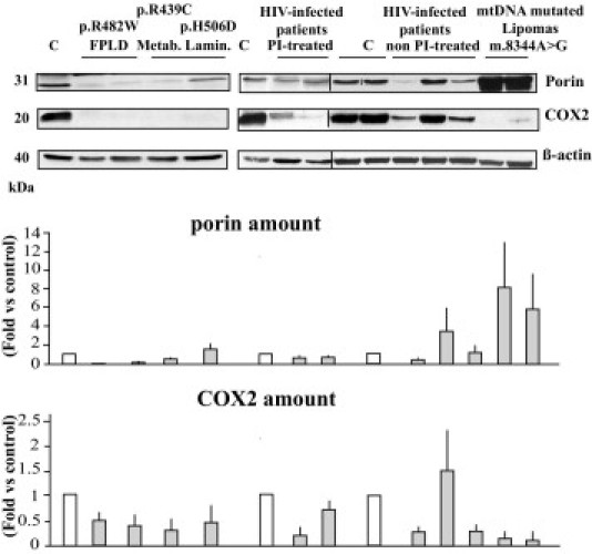
Some LMNA mutations responsible for insulin-resistant lipodystrophic syndromes are associated with peripheral subcutaneous lipoatrophy and faciocervical fat accumulation. Their pathophysiologic characteristics are unknown. We compared histologic, immunohistologic, ultrastructural, and protein expression features of enlarged cervical subcutaneous adipose tissue (scAT) obtained during plastic surgery from four patients with LMNA p.R482W, p.R439C, or p.H506D mutations versus cervical fat from eight control subjects, buffalo humps from five patients with HIV infection treated or not with protease inhibitors, and dorsocervical lipomas from two patients with mitochondrial DNA mutations. LMNA-mutated cervical scAT and HIV-related buffalo humps were dystrophic, with an increased percentage of small adipocytes, increased fibrosis without inflammatory features, and decreased number of blood vessels, as compared with control samples. Samples from patients with LMNA mutations or protease inhibitor-based therapy demonstrated accumulation of prelamin A, altered expression of adipogenic proteins and brown fat-like features, with an increased number of mitochondria and overexpression of uncoupling protein 1 (UCP1). These features were absent in samples from control subjects and from patients with HIV not treated with protease inhibitors. Mitochondrial DNA-mutated cervical lipomas demonstrated inflammatory fibrosis with distinct mitochondrial abnormalities but neither UCP1 expression nor prelamin A accumulation. In conclusion, Enlarged cervical scAT from patients with lipodystrophy demonstrated small adipocytes, fibrosis, and decreased vessel numbers. However, only cervical fat from patients with LMNA mutations or who had received protease inhibitor therapy accumulated prelamin A and exhibited similar remodeling toward a brown-like phenotype with UCP1 overexpression and mitochondrial alterations.


Figures

Figure 1
Figure 1
Cervical fat from patients with LMNA- or HIV-related lipodystrophies or with mitochondrial lipomatosis demonstrates a dystrophic phenotype with fibrosis. Light microscopy analysis of cervical adipose tissue, stained with Sirius Red to detect collagen fibers. Representative photographs are shown. Adipose tissue (magnification, ×10) from patients with LMNA-linked and HIV-related lipodystrophies or with mtDNA mutated lipomas had a heterogeneous structure with clusters of small adipocytes. Note the excess of fibrosis in patient samples as compared with control samples, with bundles of fibrosis in FPLD2.
Figure 2
Figure 2
Quantification of adipocyte size and of fibrosis in cervical fat from patients and control subjects. Adipocyte size (A) and index of fibrosis (B) in cervical adipose tissue from control subjects and patients with FPLD2, metabolic laminopathies, HIV-linked lipodystrophy either protease inhibitor-treated or not, and mtDNA mutated lipomas. All results were obtained from triplicate measurements and are expressed as mean ± SEM for the control group and each patient.
Figure 3
Figure 3
Electron microscopy analysis of cervical adipose tissue from patients with LMNA-linked or HIV-related lipodystrophies or mtDNA mutations reveals intercellular fibrosis. Cervical adipose tissue from control subjects showed thin and regular cytoplasm and light intercellular areas. In contrast, thickened peripheral rims of cytoplasm (stars) and compact and thick fibrils of collagen invading the intercellular area (arrowheads) were observed in fat from patients. Large fat droplets lying free in the connective tissue (arrow) and numerous macrophages (m) were observed in mtDNA-mutated lipomas. A, adipocyte. Magnification, ×20,000, except for far right column ×5000.
Figure 4
Figure 4
Enlarged cervical fat from patients with LMNA- or HIV-related lipodystrophies does not exhibit inflammatory features. A: Tissue sections were probed using antibodies directed against CD68, a marker of proinflammatory macrophages. Note the presence of crown-like structures surrounding involutionary adipocytes (ie, lipogranulomas) in lipomas from patients with mtDNA mutations (arrows). Magnification, ×20. B: Quantitative analysis of lipogranulomas. Results were obtained from triplicate measurements and are expressed as mean ± SEM for each subject. The number of lipogranulomas was high in fat from mtDNA-mutated lipomas but low in cervical fat from control subjects and patients with LMNA mutations or HIV infection.
Figure 5
Figure 5
Patient cervical fat demonstrates a decreased number of vessels as compared with fat from control subjects. A: Tissue sections were probed using antibodies directed against CD34, a marker of endothelial cells. Magnification, ×20. B: Quantification of α-SMA (a marker of the media layer of arteries) and CD34-stained surfaces in samples from control subjects and from patients with LMNA mutations (blue), HIV infection (green), or mtDNA mutations (pink).
Figure 6
Figure 6
Cervical adipose tissue from patients with LMNA mutations or HIV infection currently treated using protease inhibitors demonstrates altered adipogenic factors and increased UCP1 expression with prelamin A accumulation. Adipose tissue lysates were submitted to Western blot analysis using antibodies directed against SREBP-1, PPARγ, prelamin A, UCP1, or β-actin (used as an index of total protein expression). Representative blots from triplicate experiments are shown, and quantifications are expressed as fold changes versus control values. The accumulation of prelamin A is associated with increased expression of UCP1 in enlarged cervical fat from patients with LMNA mutations or HIV infection treated using a protease inhibitor–based therapeutic regimen. C, control subject fat samples.
Figure 7
Figure 7
Cervical adipose tissue from patients with LMNA mutations or protease inhibitor–based treated HIV infection demonstrates an increased number of mitochondria. Electron microscopy analysis (magnification, ×20,000) (A) and immunohistochemical analysis using antibodies directed against mitochondria (magnification, ×40) (B) in cervical adipose tissue samples. Increased density of mitochondria (arrows) was observed in all patient samples, but was maximal in mtDNA-mutated fat.
Figure 8
Figure 8
Patient cervical fat demonstrates alterations of mitochondrial protein expression. Adipose tissue lysates were submitted to Western blot analysis using antibodies directed against the mitochondrial proteins VDAC/porin, COX2 (mtDNA-encoded), and β-actin, used as an index of total protein expression. Representative blots from triplicate experiments are shown, and quantifications are expressed as fold changes versus control values. C, control subject fat samples.

References

    1. Mattout A., Dechat T., Adam S.A., Goldman R.D., Gruenbaum Y. Nuclear lamins, diseases and aging. Curr Opin Cell Biol. 2006;18:335–341. - PubMed
    1. Cao H., Hegele R.A. Nuclear lamin A/C R482Q mutation in Canadian kindreds with Dunnigan-type familial partial lipodystrophy. Hum Mol Genet. 2000;9:109–112. - PubMed
    1. Shackleton S., Lloyd D.J., Jackson S.N., Evans R., Niermeijer M.F., Singh B.M., Schmidt H., Brabant G., Kumar S., Durrington P.N., Gregory S., O'Rahilly S., Trembath R.C. LMNA, encoding lamin A/C, is mutated in partial lipodystrophy. Nat Genet. 2000;24:153–156. - PubMed
    1. Vigouroux C., Caron-Debarle M., Le Dour C., Magré J., Capeau J. Molecular mechanisms of human lipodystrophies: from adipocyte lipid droplet to oxidative stress and lipotoxicity. Int J Biochem Cell Biol. 2011;43:862–876. - PubMed
    1. Garg A., Agarwal A.K. Lipodystrophies: disorders of adipose tissue biology. Biochim Biophys Acta. 2009;1791:507–513. - PMC - PubMed

Publication types