. 2011 Nov;179(5):2266-77.
doi: 10.1016/j.ajpath.2011.07.040. Epub 2011 Sep 21.

VEGFR-1 mediates endothelial differentiation and formation of blood vessels in a murine model of infantile hemangioma

Affiliations

VEGFR-1 mediates endothelial differentiation and formation of blood vessels in a murine model of infantile hemangioma

Elisa Boscolo et al. Am J Pathol. 2011 Nov.

Abstract

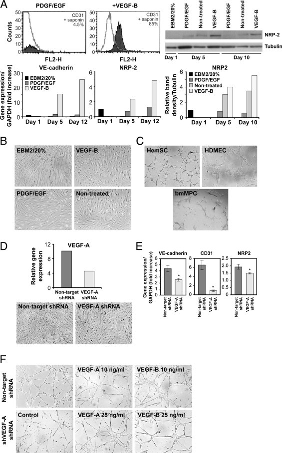
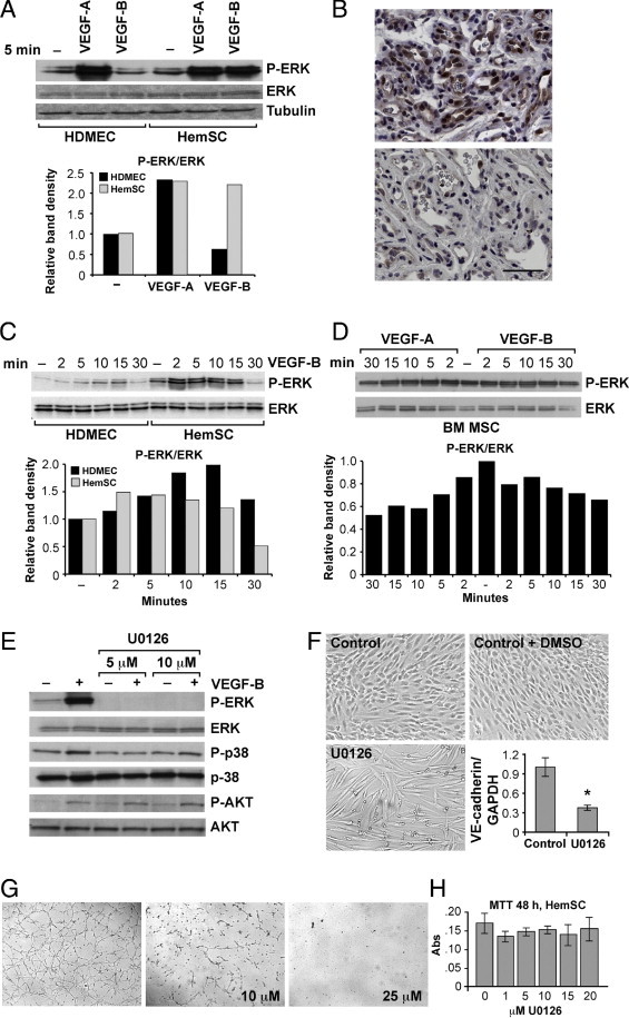
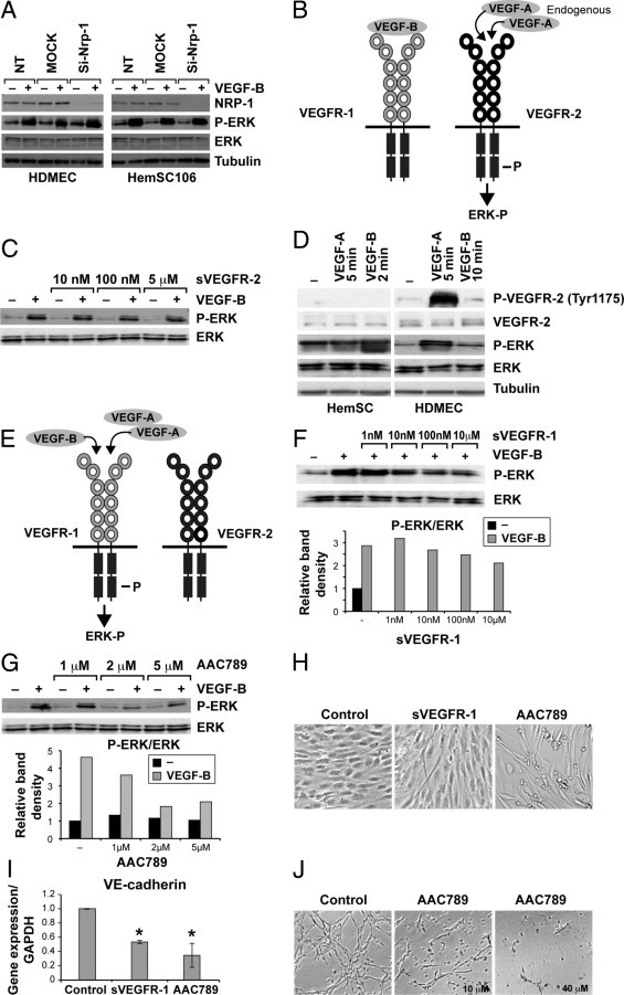
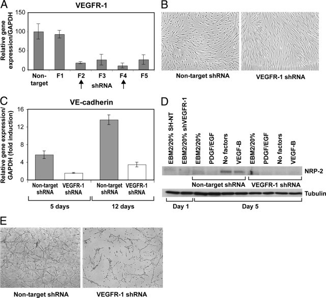
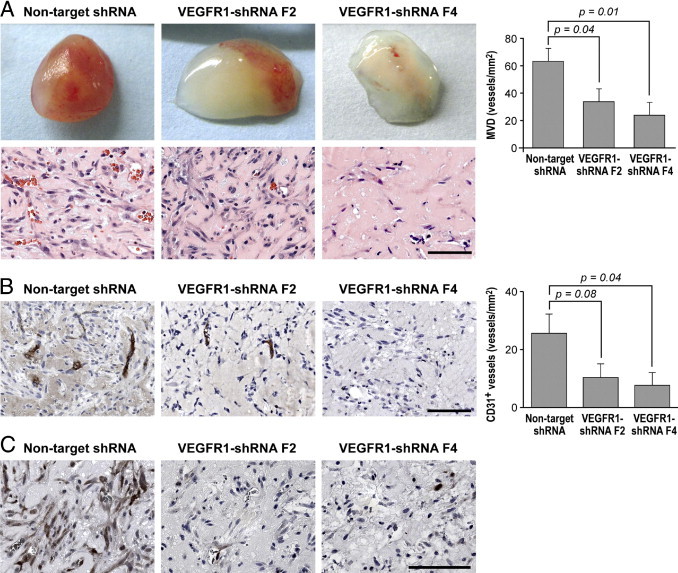
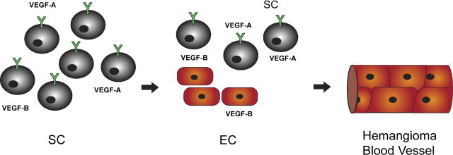
Vascular endothelial growth factor receptor-1 (VEGFR-1) is a member of the VEGFR family, and binds to VEGF-A, VEGF-B, and placental growth factor. VEGFR-1 contributes to tumor growth and metastasis, but its role in the pathological formation of blood vessels is still poorly understood. Herein, we used infantile hemangioma (IH), the most common tumor of infancy, as a means to study VEGFR-1 activation in pathological vasculogenesis. IH arises from stem cells (HemSCs) that can form the three most prominent cell types in the tumor: endothelial cells, pericytes, and adipocytes. HemSCs can recapitulate the IH life cycle when injected in immuncompromised mice, and are targeted by corticosteroids, the traditional treatment for IH. We report here that VEGF-A or VEGF-B induces VEGFR-1-mediated ERK1/2 phosphorylation in HemSCs and promotes differentiation of HemSCs to endothelial cells. Studies of VEGFR-2 phosphorylation status and down-regulation of neuropilin-1 in the HemSCs demonstrate that VEGFR-2 and NRP1 are not needed for VEGF-A- or VEGF-B-induced ERK1/2 activation. U0216-mediated blockade of ERK1/2 phosphorylation or shRNA-mediated suppression of VEGFR-1 prevents HemSC-to-EC differentiation. Furthermore, the down-regulation of VEGFR-1 in the HemSCs results in decreased formation of blood vessels in vivo and reduced ERK1/2 activation. Thus, our study reveals a critical role for VEGFR-1 in the HemSC-to-EC differentiation that underpins pathological vasculogenesis in IH.


Figures

Figure 1
Figure 1
VEGF-B and endogenous VEGF-A induce HemSC-to-EC differentiation. A: Histogram (shaded gray) represents CD31 expression in HemSCs after 8 days of exposure to PDGF/EGF (both at 10 ng/mL) or VEGF-B (10 ng/mL) in serum-free medium; gray line, matching IgG control (top left). Immunoblot shows NRP-2 in HemSCs at day 1 in EBM2/20% FBS (EBM2/20%), and after 5 and 10 days in endothelial differentiation medium with PDGF-BB plus EGF (each at 10 ng/mL), or no treatment or VEGF-B (10 ng/mL). Tubulin is shown as a loading control (top right). Relative immunoblot band intensities (NRP2/Tubulin) shown in the graph below. On the bottom left, RT-PCR for VE-cadherin and NRP-2 HemSCs in EBM2/20%, in endothelial differentiation medium with PDGF plus EGF, or VEGF-B, at days 1, 5 and 12, normalized to day 1 in EBM2/20%. B: HemSCs cultured for 8 days in EBM2/20% or endothelial differentiation medium with addition of VEGF-B, PDGF/EGF, or without factors (nontreated). C: HemSCs, HDMECs, and bmMPCs plated on Matrigel for 18 hours. D: RT-PCR for VEGF-A in HemSCs infected with lentivirus containing nontarget shRNA and VEGF-A shRNA (top). Cells cultured for 5 days in endothelial differentiation medium, and (E) RT-PCR shows levels of VE-cadherin, CD31, and NRP-2. F: Nontarget and VEGF-A shRNA HemSCs cultured on Matrigel for 18 hours. In four right panels, tube formation assessed in response to exogenous VEGF-A and VEGF-B (10 and 25 ng/mL).
Figure 2
Figure 2
VEGF-B and VEGF-A activate ERK phosphorylation in HemSCs. Immunoblot of phospho-ERK (P-ERK) and ERK in HDMECs and HemSCs in response to (A) 5 minutes' exposure to VEGF-A (25 ng/mL) or VEGF-B (10 ng/mL). Relative immunoblot band intensities (phosphoERK/ERK) are shown in the graph. B: Proliferating (top panel) and involuting (bottom panel) IH specimen stained with anti-phosphoERK antibody. Scale bar = 20 μm. C: Immunoblot and graphs of relative band intensities of phospho-ERK and ERK in HDMECs and HemSCs in response to VEGF-B (25 ng/mL) at different time points, and (D) in bone marrow mesenchymal stem cells (bmMSC) after treatment with VEGF-A or VEGF-B (both at 25 ng/mL). E: HemSCs pretreated with U0126 (5 and 10 μmol/L) for 1 hour and incubated for 2 minutes with VEGF-B (25 ng/mL) subjected to immunoblot analysis for phospho-ERK, ERK, phosphor-p38, p38, and phosphoAKT, AKT. F: HemSCs cultured for 5 days in endothelial differentiation medium (Control), or with DMSO (Control+DMSO) or U0126, and RT-PCR analysis of VE-cadherin in control (DMSO) and U0126 (10 μmol/L) treated HemSCs. G: Tube formation in Matrigel assessed for HemSCs at 18 hours after control (DMSO) and U0126 (10 μmol/L and 25 μmol/L) treatment. H: Cell survival, measured with MTT assay, for HemSCs exposed to different concentrations of U0126. Data expressed as means ± SD.
Figure 3
Figure 3
NRP1 and VEGFR-2 are not involved in HemSC-to EC differentiation. HDMECs and HemSCs subjected to siRNA to down-regulate NRP1 and immunoblot: A: NRP1, phosphoERK, and ERK levels in cells treated or not treated with VEGF-B (25 ng/mL); tubulin is loading control. B: Schematic of hypothesis of indirect role for VEGFR-1 in the HemSC-to-EC differentiation, as described in text. C: Immunoblot of phosphoERK and ERK in HemSCs pre-treated for 1 hour with soluble VEGFR-2 (sVEGFR-2) followed by stimulation with VEGF-B (25 ng/mL). D: HemSCs and HDMECs incubated with VEGF-A (25 ng/mL, 5 minutes) or VEGF-B (25 ng/mL, 2 minutes for HemSCs and 10 minutes for HDMECs). Cell lysates analyzed for phosphoVEGFR-2 (Tyr1175), total VEGFR-2, phosphoERK, ERK, and tubulin (loading control). E: Schematic of hypothesis of direct role for VEGFR-1 in the HemSC-to-EC differentiation, as described in in text. F: Immunoblot and graphs of relative band intensities for phosphoERK and ERK in HemSCs stimulated for 2 minutes with VEGF-B (25 ng/mL) after 1 hour of pretreatment with soluble VEGFR-1 (sVEGFR-1) (1 nmol to 10 μmol/L), or (G) with AAC789 (1–5 μmol/L). H: HemSCs cultured for 5 days in serum-free medium plus VEGF-B alone (Control), or VEGF-B plus sVEGFR-1 (5 μg/mL) or AAC789 (5 μmol/L), and (I) quantitative RT-PCR analysis of VE-cadherin in control, sVEGFR-1, and AAC789 treated HemSCs. J: Tube formation in Matrigel in HemSCs 18 hours after control (DMSO) and AAC789 (10 μmol/L and 40 μmol/L) treatment. Data are mean ± SD.
Figure 4
Figure 4
Silencing of VEGFR-1 prevents HemSC-to-EC differentiation in vitro. A: RT-PCR shows VEGFR-1 expression levels in HemSCs after infection with lentivirus encoding five different shRNA sequences (F1–F5) targeting VEGFR-1, and control nontarget sequence. B: Non-target and VEGFR-1 shRNA (F4) HemSCs cultured for 5 days in endothelial differentiation medium, and (C) quantitative RT-PCR illustrates relative VE-cadherin expression after 5 and 12 days in culture. D: Immunoblot for NRP-2 in HemSCs, nontarget and VEGFR-1 shRNA at days 1 and 5 after culture in regular growth medium (EBM2/20%) or in serum-free medium plus PDGF and EGF (10 ng/mL each), no factors, or VEGF-B (10 ng/mL); tubulin was used as loading control. E: Nontarget and VEGFR-1 shRNA HemSCs cultured in Matrigel thin layer for 18 hours. Data expressed as means ± SD.
Figure 5
Figure 5
Silencing of VEGFR-1 prevents formation of blood vessels in vivo. A: Representative photographs of Matrigel explants at day 7 after injection of HemSCs nontarget and VEGFR-1 shRNA (F2 and F4), corresponding sections stained for H&E, and quantification of total microvessel density (MVD) as microvessels/mm2. B: Anti-human-CD31 staining in Matrigel explants from experiment in A, graph shows quantification of human CD31+ vessels. C: PhosphoERK1/2 staining in Matrigel explants from experiment in A. Scale bar = 100 μm. Data are mean ± SEM.
Figure 6
Figure 6
Hypothesis of HemSC-to-EC differentiation in IH tissue. HemSCs produce VEGF-A, which in turn mediates endothelial differentiation in an autocrine fashion by binding to VEGFR1. HemSC-derived ECs secrete VEGF-B, which can then potentiate further HemSC-to-EC differentiation leading to IH blood vessel formation.

Similar articles

Cited by

References

    1. Fong G.H., Rossant J., Gertsenstein M., Breitman M.L. Role of the Flt-1 receptor tyrosine kinase in regulating the assembly of vascular endothelium. Nature. 1995;376:66–70. - PubMed
    1. Hiratsuka S., Maru Y., Okada A., Seiki M., Noda T., Shibuya M. Involvement of Flt-1 tyrosine kinase (vascular endothelial growth factor receptor-1) in pathological angiogenesis. Cancer Res. 2001;61:1207–1213. - PubMed
    1. Kaplan R.N., Riba R.D., Zacharoulis S., Bramley A.H., Vincent L., Costa C., MacDonald D.D., Jin D.K., Shido K., Kerns S.A., Zhu Z., Hicklin D., Wu Y., Port J.L., Altorki N., Port E.R., Ruggero D., Shmelkov S.V., Jensen K.K., Rafii S., Lyden D. VEGFR1-positive haematopoietic bone marrow progenitors initiate the pre-metastatic niche. Nature. 2005;438:820–827. - PMC - PubMed
    1. Fischer C., Mazzone M., Jonckx B., Carmeliet P. FLT1 and its ligands VEGFB and PlGF: drug targets for anti-angiogenic therapy? Nat Rev Cancer. 2008;8:942–956. - PubMed
    1. Carmeliet P., Ferreira V., Breier G., Pollefeyt S., Kieckens L., Gertsenstein M., Fahrig M., Vandenhoeck A., Harpal K., Eberhardt C., Declercq C., Pawling J., Moons L., Collen D., Risau W., Nagy A. Abnormal blood vessel development and lethality in embryos lacking a single VEGF allele. Nature. 1996;380:435–439. - PubMed

Publication types

MeSH terms

Substances