. 2012 Jan;45(1):425-37.
doi: 10.1016/j.nbd.2011.09.002. Epub 2011 Sep 13.

Activation of glycogen synthase kinase-3 beta mediates β-amyloid induced neuritic damage in Alzheimer's disease

Affiliations

Activation of glycogen synthase kinase-3 beta mediates β-amyloid induced neuritic damage in Alzheimer's disease

B DaRocha-Souto et al. Neurobiol Dis. 2012 Jan.

Abstract

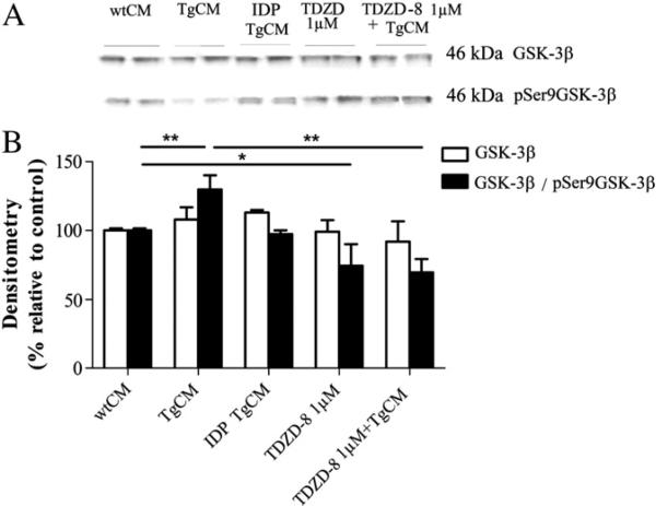
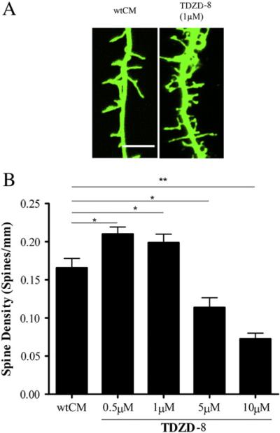
β-Amyloid (Aβ) plaques in Alzheimer (AD) brains are surrounded by severe dendritic and axonal changes, including local spine loss, axonal swellings and distorted neurite trajectories. Whether and how plaques induce these neuropil abnormalities remains unknown. We tested the hypothesis that oligomeric assemblies of Aβ, seen in the periphery of plaques, mediate the neurodegenerative phenotype of AD by triggering activation of the enzyme GSK-3β, which in turn appears to inhibit a transcriptional program mediated by CREB. We detect increased activity of GSK-3β after exposure to oligomeric Aβ in neurons in culture, in the brain of double transgenic APP/tau mice and in AD brains. Activation of GSK-3β, even in the absence of Aβ, is sufficient to produce a phenocopy of Aβ-induced dendritic spine loss in neurons in culture, while pharmacological inhibition of GSK-3β prevents spine loss and increases expression of CREB-target genes like BDNF. Of note, in transgenic mice GSK-3β inhibition ameliorated plaque-related neuritic changes and increased CREB-mediated gene expression. Moreover, GSK-3β inhibition robustly decreased the oligomeric Aβ load in the mouse brain. All these findings support the idea that GSK3β is aberrantly activated by the presence of Aβ, and contributes, at least in part, to the neuronal anatomical derangement associated with Aβ plaques in AD brains and to Aβ pathology itself.


Figures

Fig. 1
Fig. 1
(A, B) 24 hour exposure of neurons to Tg2576 CM resulted in a significant reduction of spine density when compared to neurons exposed to wt media. Exposure to oligomeric Aβ isolated on the SEC column from AD brains induced a similar reduction in the number of dendritic spines, and immunodepletion using 3D6 (=IDPTgCM) antibody completely rescued this phenotype. Data are derived from 20 neurons per condition. Calibration bars 10 μm. *** p<0.001.
Fig. 2
Fig. 2
(A, B). 24 hour exposure of neurons to Tg2576 CM significantly decreased the proportion of thin and mushroom spines. TDZD-8 treatment at 1 μM increased the overall amount of dendritic spines by over 25%, restored the proportion of thin and mushroom spines to control levels, and increased the relative proportion of stubby spines. Transfection of neurons with a wt GKS-3β isoform significantly decreased the amount of all three classes of spines, thin, mushroom and stubby in the absence of Aβ. Data are derived from 20 neurons per condition. *p<0.05 ** p<0.01 *** p<0.001.
Fig. 3
Fig. 3
(A, B). Total level of GSK-3β in nuclear extracts did not significantly change in primary cultured neurons after 24 hour exposure to Tg2576 CM compared to wt CM. GSK-3β/pSer9GSK-3β ratio was increased by 30% indicating that oligomeric Aβ exposure triggers a significant increase in GSK-3β activity. 6E10 immunodepletion completely prevented the elevation of GSK-3β activity. Treatment of cultured neurons with TDZD-8 at 1 μM significantly decreased GSK-3β/pSer9GSK-3β ratio indicating that the enzyme is inhibited by this treatment. Data are derived from 24 dishes per condition. *p<0.05 ** p<0.01.
Fig. 4
Fig. 4
(A, B) Treatment of neurons with TDZD-8 at 1 μM increased the overall amount of dendritic spines by over 25% in comparison to neurons exposed to wt CM. Concentrations of TDZD-8 higher than 1 μM were toxic and resulted in significant reduction in the number of spines suggesting that excessive inhibition of GSK-3β endogenous activity might not be safe. Data are derived from 20 neurons per condition. Calibration bars 10 μm. *p<0.05 ** p<0.01.
Fig. 5
Fig. 5
(A, B) TDZD-8 at 1 μM fully rescued spine loss triggered by exposure to oligomeric Aβ containing Tg2576 CM. 14 day transfection of wt GSK-3β into neurons led to a very robust spine loss recapitulating that observed after exposure to Tg2576 CM. The phenotype is even more dramatic after transfection of a constitutively active mutant form of GSK-3β (GSK-3β mut (S9A)) that leads to a complete loss of all dendritic spines in cultured neurons. Data are derived from 20 neurons per condition. Calibration bars 10 μm. *p<0.05 ** p<0.01 ***p<0.001.
Fig. 6
Fig. 6
(A–D) GSK-3β levels in total homogenates (A, B) and nuclear fractions (C, D) prepared from the temporal cortex of AD samples were markedly decreased compared to those seen in control samples, as expected due to neuronal cell loss in AD. The ratio GSK-3β/pSer9GSK-3β was increased in the total homogenates from AD brains by 260%, and by 450% in the nuclear fraction when compared to controls indicating that GSK-3β is more active in AD brains (n=12 brains per group). *p<0.05 ** p<0.01.
Fig. 7
Fig. 7
(A) The total amount of Nab61 reactive oligomeric Aβ deposits was decreased in mice treated with NP12 in comparison to those treated with vehicle. (B) The regional exam further confirmed the treatment effect on Nab61 accumulation in the cingulated cortex and EC (n=6 animals per group). (C) Representative photomicrographs of sections immunostained with Nab61 through Cg, CA1 and EC from NP12 and vehicle treated animals. Calibration bars 200 μm. EC = entorhinal cortex, Cg = cingulate. *p<0.05 ** p<0.01.
Fig. 8
Fig. 8
(A) Axons far (>50 μm) from plaques were significantly curvier in 15 month-old APPsw–tauvlw mice compared to non-Tg littermate controls. Treatment with NP12 restored the normal straight pattern of axons close to plaques (<50 μm) and far from them (n=90–135 axons analyzed per group). (B) Representative image of SMI312-positive neurites immunostaining (red) and senile plaque (Thioflavine S) (green). Curvature ratio was calculated by dividing the curvilinear length of the axon by the straight line length of the axon. (C) Neuritic dystrophies (size of dystrophies defined as areas of swelling >2.5 μm in diameter in neurites) can be seen in the brain of APPsw–tauvlw mice at 15 months of age in association with amyloid deposits. NP12 treatment significantly decreased the amount of dystrophies near plaques in the brain of APPsw–tauvlw compared to vehicle treated mice. (D) Representative image of SMI312-positive neurities immunostaining (red) and senile plaque (Thioflavine S) (green). Data are derived from 6 animals per group. Calibration bar 25 μm. **p<0.01 ***p<0.001.
Fig. 9
Fig. 9
(A–H) Increased activity of GSK-3β triggered by exposure of neurons to Tg2576 CM media resulted in a significant increase in the inhibitory phosphorylation at serine 129 of CREB promoted by this enzyme and a significant decrease in the amount of BDNF. Treatment with TDZD-8 at 1 μm concentration significantly decreased the inhibitory phosphorylation at Ser 129 of CREB mediated by GSK-3β and restored the normal level of BDNF in neuronal cultures. Data are derived from 24 dishes per condition. *p<0.05 ** p<0.01 ***p<0.001.
Fig. 10
Fig. 10
(A–H) A significant increase in GKS-3β mediated inhibitory phosphorylation at serine 129 of CREB and concordant significant decreases in the amount of c-fos and ARC were observed in brain extracts from 15 month-old APPsw–tauvlw mice in comparison to similarly aged wt mice. These changes were fully rescued by pharmacological inhibition of GSK-3β in APPsw–tauvlw mice. Data are derived from n=3 animals per group. *p<0.05 ** p<0.01 ***p<0.001.
Fig. 11
Fig. 11
(A–H) A significant increase in the inhibitory phosphorylation at serine 129 of CREB and a decrease in the levels of ARC and BDNF were observed in the temporal cortex of AD brains in comparison to cognitively normal controls. Data are derived from n=11–12 brains per group. *p<0.05 ***p<0.001.

References

    1. Akiyama H, et al. Pin1 promotes production of Alzheimer's amyloid beta from beta-cleaved amyloid precursor protein. Biochem. Biophys. Res. Commun. 2005;336:521–529. - PubMed
    1. Aplin AE, et al. Effect of increased glycogen synthase kinase-3 activity upon the maturation of the amyloid precursor protein in transfected cells. Neuroreport. 1997;8:639–643. - PubMed
    1. Bao Y, et al. Activation of peroxisome proliferator-activated receptor gamma inhibits endothelin-1-induced cardiac hypertrophy via the calcineurin/NFAT signaling pathway. Mol. Cell. Biochem. 2008;317:189–196. - PubMed
    1. Barco A, et al. Expression of constitutively active CREB protein facilitates the late phase of long-term potentiation by enhancing synaptic capture. Cell. 2002;108:689–703. - PubMed
    1. Beurel E, Jope RS. The paradoxical pro- and anti-apoptotic actions of GSK3 in the intrinsic and extrinsic apoptosis signaling pathways. Prog. Neurobiol. 2006;79:173–189. - PMC - PubMed

Publication types

MeSH terms