

Executive function in chronic pain patients and healthy controls: different cortical activation during response inhibition in fibromyalgia
- PMID: 21945593
- PMCID: PMC3715316
- DOI: 10.1016/j.jpain.2011.06.007
Executive function in chronic pain patients and healthy controls: different cortical activation during response inhibition in fibromyalgia
Abstract
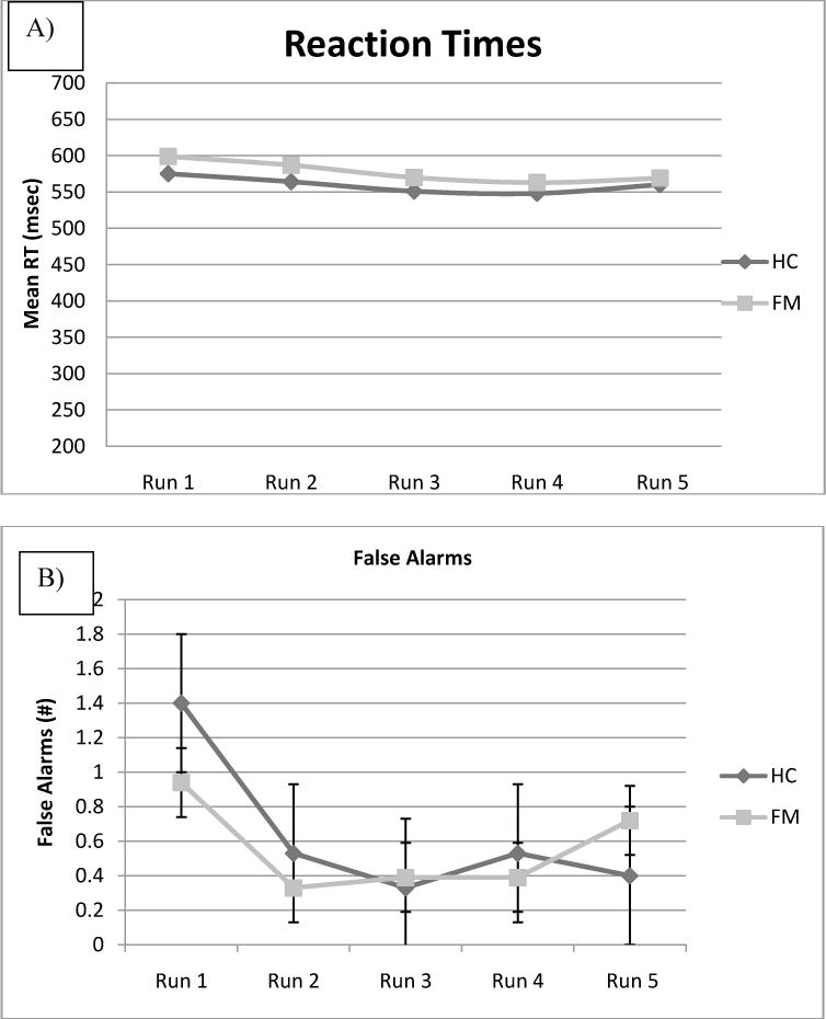
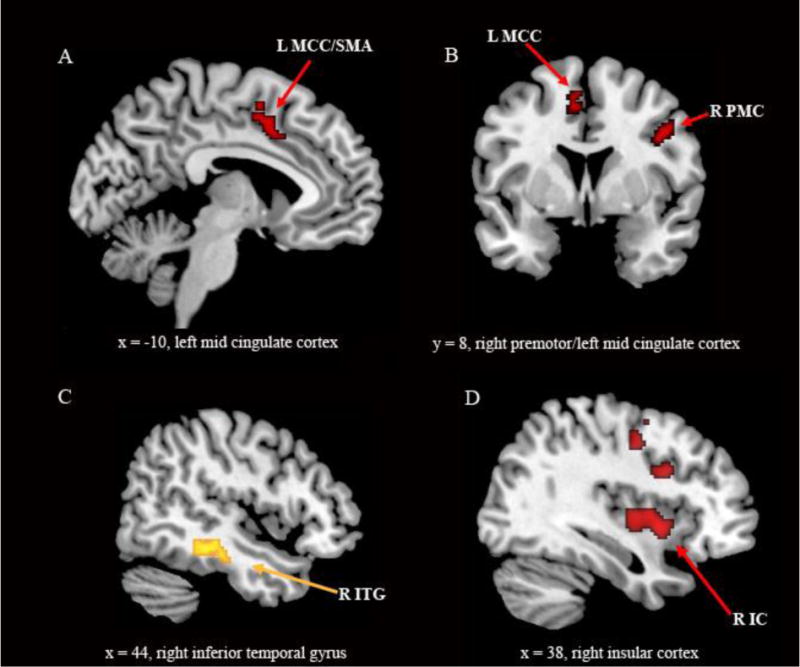
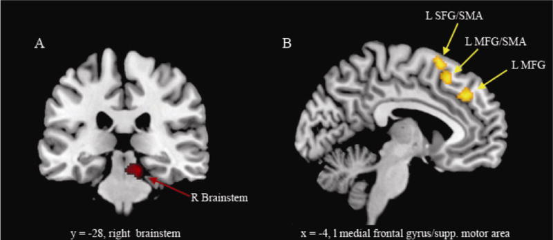
The primary symptom of fibromyalgia (FM) is chronic, widespread pain; however, patients report additional symptoms including decreased concentration and memory. Performance-based deficits are seen mainly in tests of working memory and executive function. Neural correlates of executive function were investigated in 18 FM patients and 14 age-matched healthy controls during a simple Go/No-Go task (response inhibition) while they underwent functional magnetic resonance imaging (fMRI). Performance was not different between FM and healthy control, in either reaction time or accuracy. However, fMRI revealed that FM patients had lower activation in the right premotor cortex, supplementary motor area, midcingulate cortex, putamen and, after controlling for anxiety, in the right insular cortex and right inferior frontal gyrus. A hyperactivation in FM patients was seen in the right inferior temporal gyrus/fusiform gyrus. Despite the same reaction times and accuracy, FM patients show less brain activation in cortical structures in the inhibition network (specifically in areas involved in response selection/motor preparation) and the attention network along with increased activation in brain areas not normally part of the inhibition network. We hypothesize that response inhibition and pain perception may rely on partially overlapping networks, and that in chronic pain patients, resources taken up by pain processing may not be available for executive functioning tasks such as response inhibition. Compensatory cortical plasticity may be required to achieve performance on a par with control groups.
Perspective: Neural activation (fMRI) during response inhibition was measured in fibromyalgia patients and controls. FM patients show lower activation in the inhibition and attention networks and increased activation in other areas. Inhibition and pain perception may use overlapping networks: resources taken up by pain processing may be unavailable for other processes.
Copyright © 2011 American Pain Society. Published by Elsevier Inc. All rights reserved.
Figures
References
-
- Apkarian AV, Bushnell MC, Treede RD, Zubieta JK. Human brain mechanisms of pain perception and regulation in health and disease. Eur J Pain. 2005;9:463–484. - PubMed
-
- Bingel U, Tracey I. Imaging CNS modulation of pain in humans. Physiology (Bethesda) 2008;23:371–380. - PubMed
-
- Bradley LA, McKendree-Smith NL, Alberts KR, Alarcon GS, Mountz JM, Deutsch G. Use of neuroimaging to understand abnormal pain sensitivity in fibromyalgia. Curr Rheumatol Rep. 2000;2:141–148. - PubMed
-
- Cappelleri JC, Bushmakin AG, McDermott AM, Dukes E, Sadosky A, Petrie CD, Martin S. Measurement properties of the Medical Outcomes Study Sleep Scale in patients with fibromyalgia. Sleep Med. 2009;10:766–770. - PubMed
-
- Caseras X, Mataix-Cols D, Giampietro V, Rimes KA, Brammer M, Zelaya F, Chalder T, Godfrey EL. Probing the working memory system in chronic fatigue syndrome: a functional magnetic resonance imaging study using the n-back task. Psychosom Med. 2006;68:947–955. - PubMed
Publication types
MeSH terms
Grants and funding
LinkOut - more resources
Full Text Sources
Medical
