. 2011 Nov;179(5):2315-26.
doi: 10.1016/j.ajpath.2011.07.038. Epub 2011 Sep 23.

Substance P induces CCN1 expression via histone deacetylase activity in human colonic epithelial cells

Affiliations

Substance P induces CCN1 expression via histone deacetylase activity in human colonic epithelial cells

Hon Wai Koon et al. Am J Pathol. 2011 Nov.

Abstract

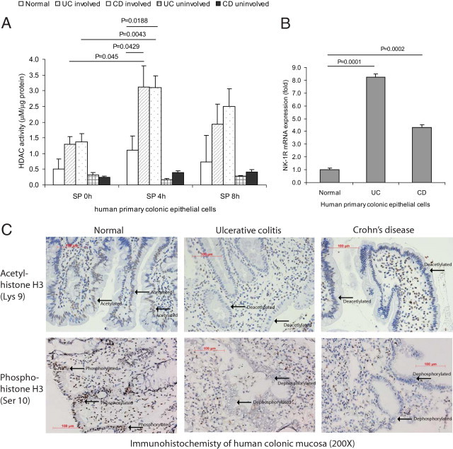
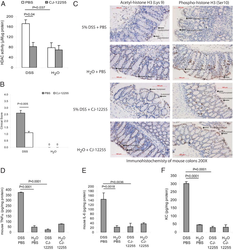
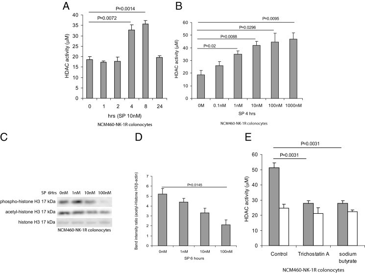
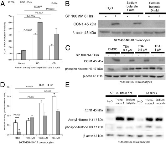
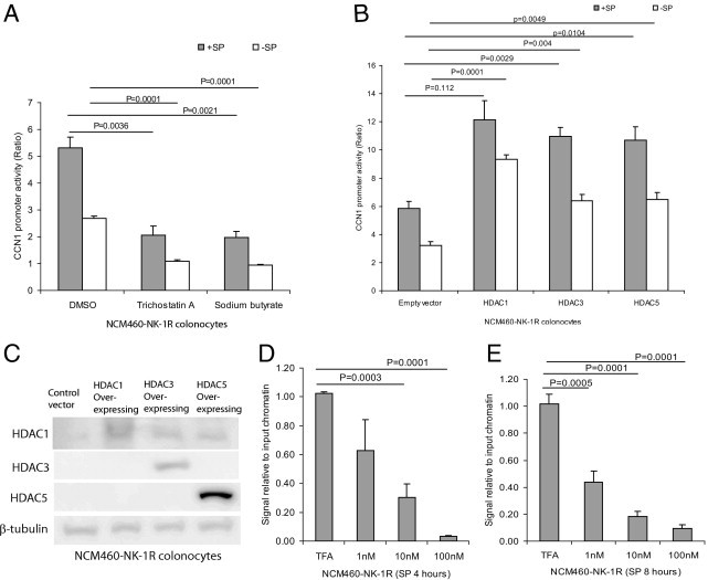
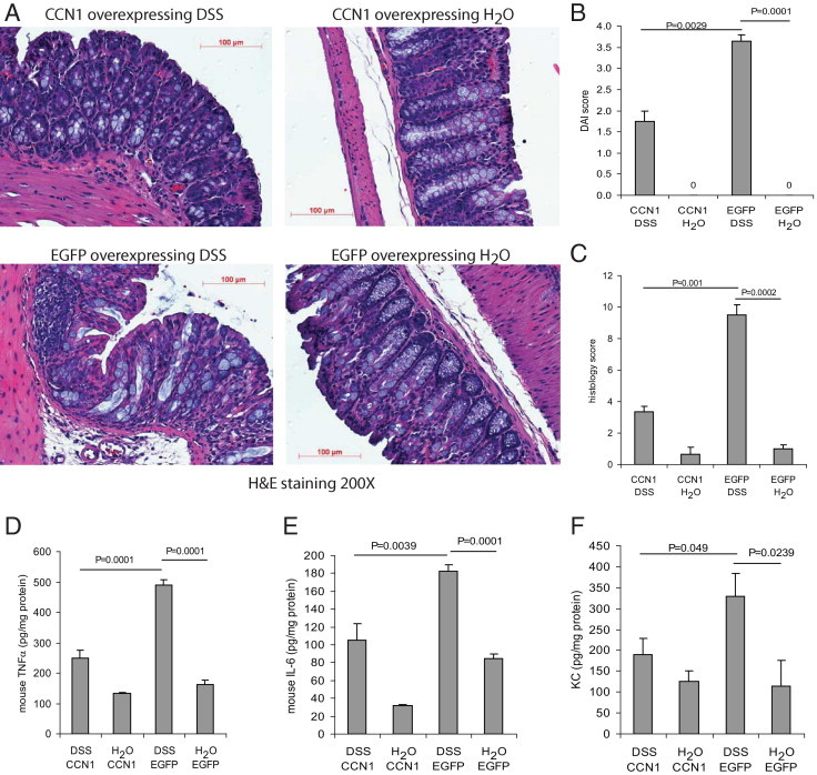
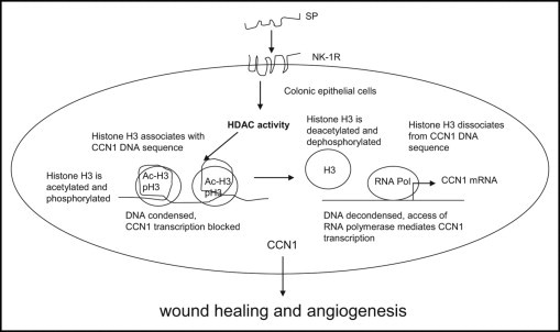
We have shown that substance P (SP) and its neurokinin-1 receptor (NK-1R) regulate intestinal angiogenesis by increasing expression of protein CYR61 (the cysteine-rich angiogenic inducer 61, or CCN1) in colonic epithelial cells. However, the mechanism involved in SP-induced CCN1 expression has not been studied, and the outcome of increased CCN1 expression in the development of colitis is not fully understood. Because histone deacetylase (HDAC) modulates transcription of several genes involved in inflammation, we investigated participation of HDAC in SP-induced CCN1 expression in human colonic epithelial NCM460 cells overexpressing NK-1R (NCM460-NK-1R) and in primary colonocytes. SP increased HDAC activity with deacetylation and dephosphorylation of nucleosome protein histone H3 in NCM460-NK-1R and/or primary colonocytes. Histone deacetylation and dephosphorylation was observed in colonic mucosa from irritable bowel disease patients. Similarly, colonic mucosal tissues from mice exposed to dextran sulfate sodium showed histone H3 deacetylation and dephosphorylation and increased HDAC activity that was reversed by the NK-1R antagonist CJ-12255. SP-induced increased CCN1 expression in NCM460-NK-1R cells was abolished by pharmacological HDAC inhibition. HDAC overexpression activated basal and SP-induced CCN1 promoter activity. Intracolonic CCN1 overexpression significantly ameliorated dextran sulfate sodium-induced colitis, with reduction of proinflammatory cytokine expression in mice. Thus, SP-mediated CCN1 expression in the inflamed human and mouse colon involves increased HDAC activity. Our results strongly suggest that increased CCN1 expression may be involved in mucosal healing during colitis.


Figures

Figure 1
Figure 1
SP mediates histone deacetylase (HDAC) activity in human primary colonic epithelial cells and colons of IBD patients. A: Human primary colonic epithelial cells from normal, involved, and uninvolved regions of IBD patients were exposed to SP (100 nmol/L) for 0 to 8 hours. HDAC activities were measured. SP significantly stimulated HDAC activity of human primary colonic epithelial cells from involved regions of UC and CD patients; HDAC activities of normal and uninvolved regions of IBD patients remained low. B: Real-time RT-PCR showed that human primary colonic epithelial cells from UC and CD patients expressed significantly more NK-1R mRNA than those from healthy subjects. C: Immunohistochemistry of colonic biopsies from healthy subjects, UC patients, and CD patients was performed. The state of acetyl-histone H3 and phospho-H3 along the mucosal lining was observed (indicated by arrows) in samples from UC and CD patients, compared with colonic samples from healthy subjects. Results are representative of six patients per group. Original magnification, ×200. Scale bars = 100 μm.
Figure 2
Figure 2
NK-1R mediates HDAC activity in colonic tissues of mice exposed to DSS. A: Mice were provided with water containing 5% DSS solution or water alone for 5 days. A separate group of mice were injected intraperitoneally with the NK-1R antagonist CJ-12255 or with PBS. HDAC activity in homogenized colonic tissues was measured. HDAC activity in inflamed colonic tissues is NK-1R-dependent. B: Clinical score of mice. Water-treated groups had no colitis, so the scores are zero. C: Acetylated and phosphorylated histone H3–positive cells were observed at the mucosa of normal colons exposed to water. Acetyl-histone H3-positive and phospho-histone H3-positive signals were reduced in inflamed colons of DSS-exposed mice, but were partially restored in DSS-exposed mice injected with NK-1R antagonist CJ-12255. This suggests that histone H3 deacetylation and dephosphorylation in colonic mucosa is NK-1R-dependent. Original magnification, ×200. Scale bars = 100 μm. D–F: Colonic levels of mouse proinflammatory cytokines TNFα (D), IL-6 (E), and KC (F). NK-1R antagonist CJ-12255 administration inhibited DSS colitis in mice. Results are representative of six mice per group.
Figure 3
Figure 3
SP induces HDAC activities in human colonic epithelial cells. A: NCM460-NK-1R colonocytes were exposed to 10 nmol/L of SP for 0 to 24 hours. B: NCM460-NK-1R colonocytes were exposed to 0 to 1000 nmol/L of SP for 4 hours. HDAC activity in cell lysates was measured. SP induces HDAC activity in colonocytes. C: NCM460-NK-1R colonocytes were exposed to 0 to 100 nmol/L of SP for 6 hours. Phospho-histone H3, acetyl-histone H3, and total histone H3 were detected by Western blot analyses. D: Quantification of acetyl-histone signal showed that SP (100 nmol/L) significantly reduced histone H3 acetylation. Results are representative of three independent experiments. SP stimulates histone H3 dephosphorylation in colonocytes. E: NCM460-NK-1R colonocytes were treated with 1 μmol/L trichostatin A or 5 mmol/L sodium butyrate for 30 minutes, followed by SP (10 nmol/L) or 0.1% trifluoroacetic acid for 8 hours. HDAC activities in cell lysates were measured. The HDAC inhibitors trichostatin A and sodium butyrate significantly inhibit SP-mediated HDAC activity. Results are representative of three independent experiments.
Figure 4
Figure 4
SP induces CCN1 expression in colonocytes via HDAC mechanism. A: Human primary colonic epithelial cells were exposed to trifluoroacetic acid or SP (100 nmol/L) for 4 hours. CCN1 mRNA levels were measured by real-time RT-PCR. Basal and SP-induced CCN1 expression levels from UC and CD patients were significantly higher than those from healthy subjects. B: NCM460-NK-1R cells were pretreated with 0 to 10 mmol/L of the HDAC inhibitor sodium butyrate for 30 minutes, followed by 100 nmol/L of SP exposure for 8 hours. Sodium butyrate treatment inhibited SP-induced CCN1 protein expression in colonocytes, as detected by Western blot analyses. C: NCM460-NK-1R cells were pretreated with 0 to 1 μmol/L of the HDAC inhibitor trichostatin A (TSA) or DMSO (vehicle) for 30 minutes, followed by 100 nmol/L of SP exposure for 8 hours. The levels of CCN1 and/or phospho-histone H3 were detected by Western blot analyses. Trichostatin A inhibits SP-induced CCN1 expression. D: Densitometric analyses of Figure 4C with phospho-histone H3 normalized to β-actin. Trichostatin A increases basal histone H3 phosphorylation and reverses SP-mediated histone H3 dephosphorylation. E: NCM460-NK-1R cells were pretreated with DMSO (vehicle), trichostatin A (1 μmol/L), or sodium butyrate (10 mmol/L) for 30 minutes, followed by 100 nmol/L of SP for 8 hours. SP-induced CCN1 expression was inhibited by both trichostatin A and sodium butyrate. Both inhibitors acetylated and phosphorylated histone H3. Results are representative of four independent experiments.
Figure 5
Figure 5
HDAC mediates CCN1 transcriptional activity in colonocytes. A: NCM460-NK-1R cells were transiently transfected with CCN1 promoter construct, followed by pretreatment with 1 μmol/L of trichostatin A, 10 mmol/L of sodium butyrate, or DMSO (vehicle) for 30 minutes and then exposure to SP (10 nmol/L) for 8 hours. CCN1 promoter activity was measured by luciferase assays. The HDAC inhibitors inhibited SP-induced CCN1 promoter activity. B: NCM460-NK-1R cells were transfected with empty vector or various isoforms of HDAC 1-, 3-, and 5-overexpressing constructs and CCN1 promoter construct, followed by exposure to SP (10 nmol/L) for 8 hours. CCN1 promoter activity was determined by luciferase assays. CCN1 promoter activity is mediated by multiple HDAC isoforms. C: HDAC 1, 3, and 5 protein expression after transient transfection of HDAC-overexpressing constructs into NCM460-NK-1R colonocytes. D and E: NCM460-NK-1R cells were exposed to SP (0 to 100 nmol/L) for 4 and 8 hours. Histone H3-DNA complex was immunoprecipitated and CCN1 DNA after immunoprecipitation was detected by real-time PCR. SP reduces association of histone H3 and CCN1 gene in colonocytes, providing access for CCN1 transcription. Results are representative of three independent experiments.
Figure 6
Figure 6
CCN1 reduces inflammation and mediates colonic healing in mice. Mice (C57BL/6, n = 4 per group) were transfected intracolonically with constructs overexpressing CCN1 and eGFP and then were treated with 5% DSS in their drinking water or water alone for 5 days. A: H&E staining shows that DSS colitis-induced tissue damage was reduced by CCN1 overexpression. Original magnification, ×200. Scale bars = 100 μm. B: Disease activity index (DAI) score and (C) histology score reflect reduction of inflammation and colonic tissue damage after CCN1 overexpression during colitis. Water-treated groups had no colitis, so the scores are zero. D–F: Colonic levels of mouse proinflammatory cytokines TNFα (D), IL-6 (E), and KC (F). CCN1 overexpression ameliorated DSS colitis and reduced colonic levels of proinflammatory cytokines. Results are representative of six mice.
Figure 7
Figure 7
Schematic of SP-mediated HDAC pathway. SP activates HDAC activity, which in turn deacetylates and possibly dephosphorylates histone H3 in colonic epithelial cells. Under normal conditions, histone H3 is associated with chromatin and blocks CCN1 transcription. On exposure to SP, histone H3 dissociates from chromatin and opens up to access of transcriptional factors and RNA polymerases. This facilitates CCN1 transcription. CCN1 mediates wound healing and angiogenesis.

References

    1. Chang M.M., Leeman S.E. Isolation of a sialogogic peptide from bovine hypothalamic tissue and its characterization as substance P. J Biol Chem. 1970;245:4784–4790. - PubMed
    1. Mantyh P.W. Neurobiology of substance P and the NK1 receptor. J Clin Psychiatry. 2002;63(Suppl 11):6–10. - PubMed
    1. Costa M., Furness J.B., Franco R., Llewellyn-Smith I., Murphy R., Beardsley A.M. Substance P in nerve tissue in the gut. Ciba Found Symp. 1982:129–144. - PubMed
    1. Maggi C.A. Capsaicin-sensitive nerves in the gastrointestinal tract. Arch Int Pharmacodyn Ther. 1990;303:157–166. - PubMed
    1. Bost K.L. Quantification of macrophage-derived substance P receptor mRNA using competitive polymerase chain reaction. Adv Exp Med Biol. 1995;373:219–223. - PubMed

Publication types

MeSH terms

Substances