. 2011 Nov;49(13):3656-69.
doi: 10.1016/j.neuropsychologia.2011.09.021. Epub 2011 Sep 19.

Age-related neural changes in autobiographical remembering and imagining

Affiliations

Age-related neural changes in autobiographical remembering and imagining

Donna Rose Addis et al. Neuropsychologia. 2011 Nov.

Abstract

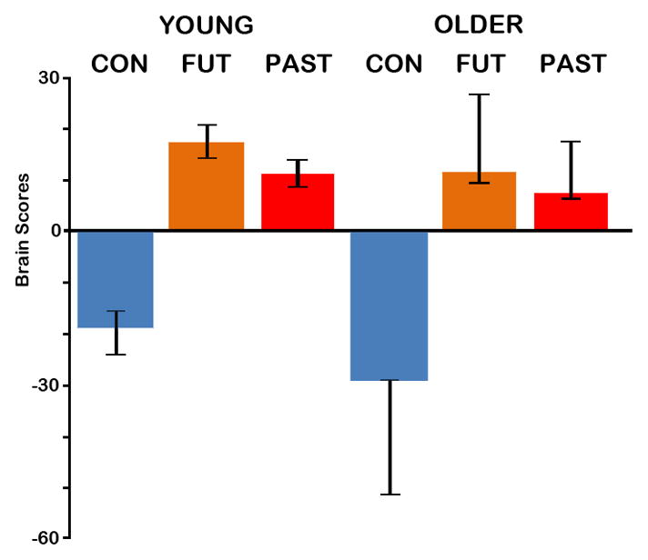
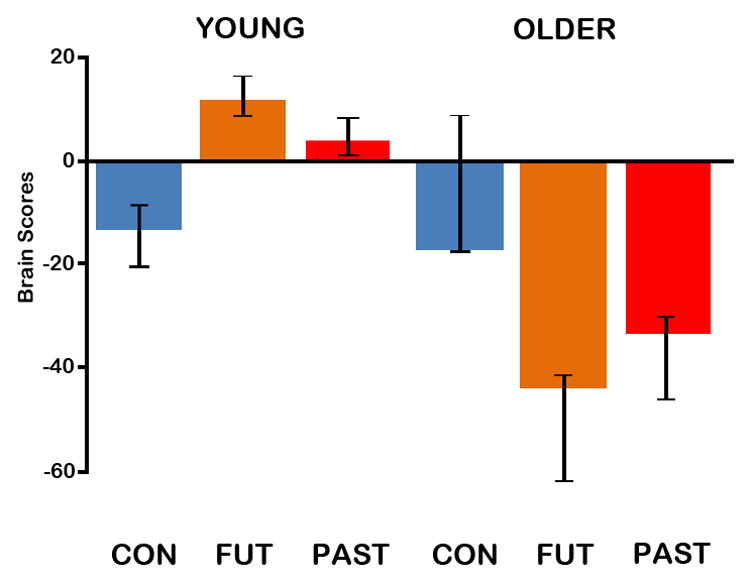
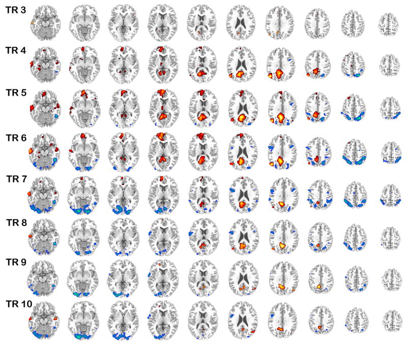
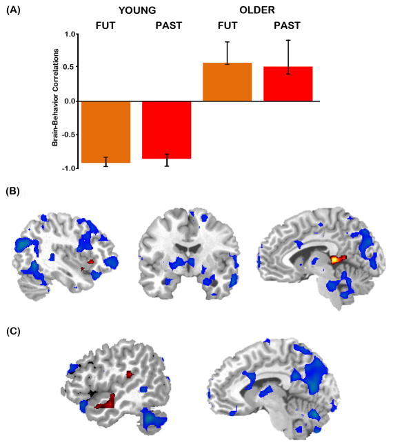
Numerous neuroimaging studies have revealed that in young adults, remembering the past and imagining the future engage a common core network. Although it has been observed that older adults engage a similar network during these tasks, it is unclear whether or not they activate this network in a similar manner to young adults. Young and older participants completed two autobiographical tasks (imagining future events and recalling past events) in addition to a semantic-visuospatial control task. Spatiotemporal Partial Least Squares analyses examined whole brain patterns of activity across both the construction and elaboration of autobiographical events. These analyses revealed that that both age groups activated a similar network during the autobiographical tasks. However, some key age-related differences in the activation of this network emerged. During the construction of autobiographical events, older adults showed less activation relative to younger adults, in regions supporting episodic detail such as the medial temporal lobes and the precuneus. Later in the trial, older adults showed differential recruitment of medial and lateral temporal regions supporting the elaboration of autobiographical events, and possibly reflecting an increased role of conceptual information when older adults describe their pasts and their futures.


Figures

Figure 1
Figure 1
The average brain scores (mean-centered across groups) with 95% confidence intervals for control and autobiographical (past and future) tasks in young and older adults associated with the condition contrast (autobiographical > control tasks). Brain scores are a weighted average of activity across all voxels associated with particular conditions. This contrast was significant (p<.001). Con=Control task; Fut=Future task; Past=Past task.
Figure 2
Figure 2
The brain regions identified by the condition contrast; activation associated with the autobiographical tasks (warm colors) and the control task (cool colors) is shown superimposed over a standard anatomical template and divided into 2 s TR (TRs 3 to 10 are shown). Images are thresholded using a bootstrap ratio of ±4 (equivalent to p < .0001).
Figure 3
Figure 3
HRF plots (percent signal change) extracted from regions identified by the condition contrast associated with (A) the autobiographical tasks (left medial parietal cortex, xyz = −4 −56 32; left medial prefrontal cortex, xyz = −12 48 −8; right hippocampus, xyz = 28 −16 −20) and (B) the control task (left superior parietal lobule, xyz = −24 −68 48; right superior parietal lobule, xyz = 32 −68 48; right inferior temporal gyrus, xyz = 52 −48 16). Images of activation are superimposed over a standard anatomical template and thresholded using a bootstrap ratio of ±4 (equivalent to p < .0001). The crosshair indicates the location of this peak voxel, and the grey bar on the plot indicates the TR from which these peak voxels were identified (TR 5). BA = Brodmann area; L = left; R = right.
Figure 4
Figure 4
The average brain scores (mean-centered across groups) with 95% confidence intervals for control and autobiographical (past and future) tasks in young and older adults associated with the interaction contrast (autobiographical > control tasks in young; control > autobiographical tasks in old). Brain scores are a weighted average of activity across all voxels associated with particular conditions. This contrast was significant (p = .05). Con=Control task; Fut=Future task; Past=Past task.
Figure 5
Figure 5
The brain regions identified by the interaction; activation associated with an autobiographical>control task effect in young but not old (warm colors) and an autobiographical>control task in old but not young (cool colors) is shown superimposed over a standard anatomical template and divided into 2 s TR (TRs 3 to 10 are shown). Images are thresholded using a bootstrap ratio of ±3 (equivalent to p < .003).
Figure 6
Figure 6
HRF plots (percent signal change) extracted from regions identified by the interaction contrast associated with (A) an autobiographical>control task effect in young but not old during TR 5 (left hippocampus, xyz = −28 −24 −12; right hippocampus, xyz = 24 −28 −12; left parahippocampal gyrus, xyz = −32 −40 −12) and (B) an autobiographical>control task effect in old than young during TR 9 (left temporal pole, xyz = −40 0 −32; right superior temporal gyrus, xyz = 52 −36 16; right hippocampus, xyz = 32 −8 −24). Images of activation are superimposed over a standard anatomical template and thresholded using a bootstrap ratio of ±3 (equivalent to p < .003). The crosshair indicates the location of this peak voxel, and the grey bar on the plot indicates the TR from which these peak voxels were identified (TR 5 or TR 9). BA = Brodmann area; L = left; R = right.
Figure 7
Figure 7
Results of the behavioral PLS analysis for detail. (A) The brain-behavior LV indicated that the regions correlated with past and future detail ratings differed significantly between young and older adults. (B) Regions in which activity correlated with detail ratings are shown in blue for young adults and red for older adults. At TR 5, regions correlated with detail in young included right lateral prefrontal and parietal cortex (left), bilateral hippocampus and right lateral temporal cortex (middle), and left medial parietal cortex and cerebellum (right). In older adults, detail correlated with insula (left) and retrosplenial cortex (right). (C) At TR 9, brain regions correlated with detail included left lateral temporal cortex for older adults and cerebellum for young adults (left) and left medial parietal cortex for young adults (right). Images of activation are superimposed over a standard anatomical template and thresholded using a bootstrap ratio of ±2 (equivalent to p < .045).

References

    1. Abraham A, Schubotz RI, von Cramon DY. Thinking about the future versus the past in personal and non-personal contexts. Brain Research. 2008;1233:106–119. - PubMed
    1. Addis DR, Cheng T, Roberts RP, Schacter DL. Hippocampal contributions to the episodic simulation of specific and general future events. Hippocampus in press. - PMC - PubMed
    1. Addis DR, McIntosh AR, Moscovitch M, Crawley AP, McAndrews MP. Characterizing spatial and temporal features of autobiographical memory retrieval networks: a partial least squares approach. Neuroimage. 2004;23:1460–1471. - PubMed
    1. Addis DR, Musicaro R, Pan L, Schacter DL. Episodic simulation of past and future events in older adults: Evidence from an experimental recombination task. Psychology and Aging. 2010;25:369–376. - PMC - PubMed
    1. Addis DR, Pan L, Vu M, Laiser N, Schacter DL. Constructive episodic simulation of the future and the past: distinct subsystems of a core brain network mediate imagining and remembering. Neuropsychologia. 2009;47:2222–2238. - PubMed

Publication types