Review
. 2012 Jan;52(1):17-30.
doi: 10.1016/j.advenzreg.2011.09.015.

Truth and consequences of sphingosine-1-phosphate lyase

Affiliations
Review

Truth and consequences of sphingosine-1-phosphate lyase

Ana Aguilar et al. Adv Biol Regul. 2012 Jan.

Abstract

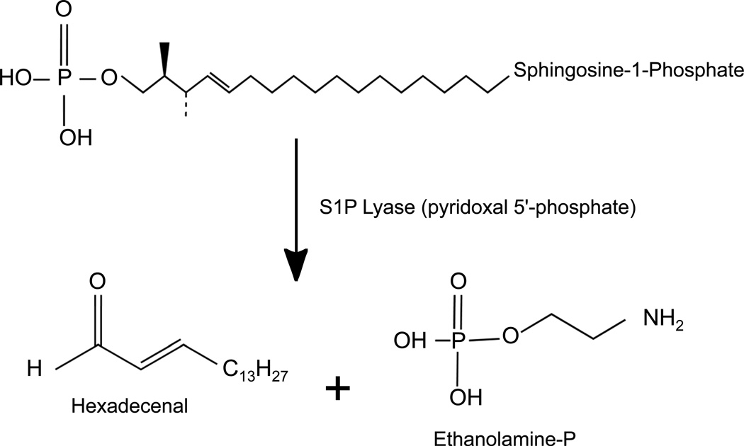
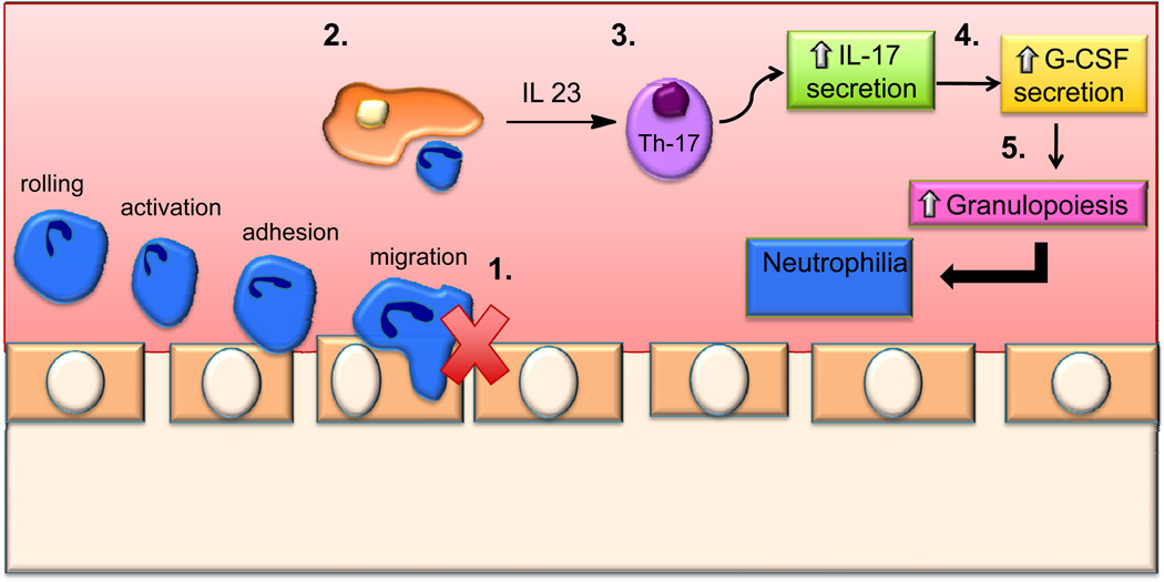
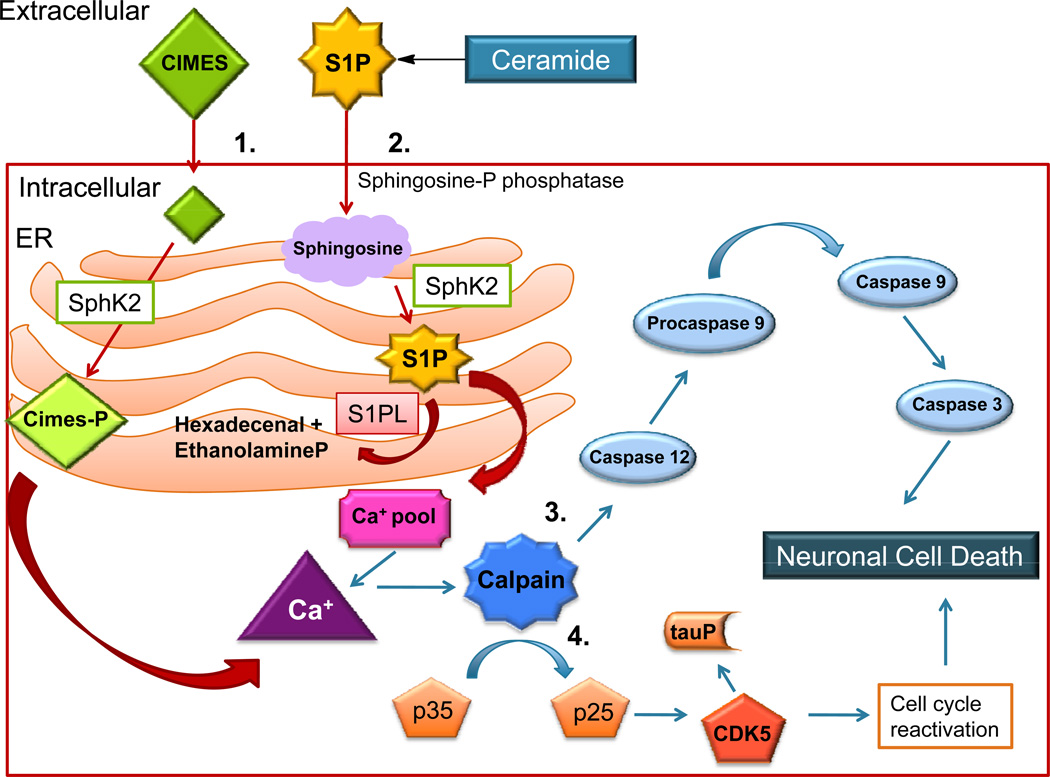
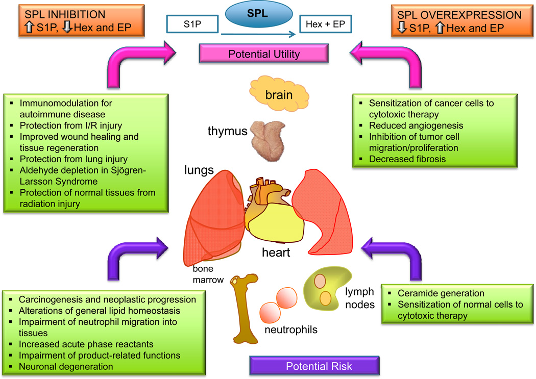
Sphingosine phosphate lyase (SPL) is an intracellular enzyme responsible for the irreversible catabolism of the lipid signaling molecule sphingosine-1-phosphate (S1P). SPL catalyzes the cleavage of S1P resulting in the formation of hexadecenal and ethanolamine phosphate. S1P functions as a ligand for a family of ubiquitously expressed G protein-coupled receptors that mediate autocrine and paracrine signals controlling cell migration, proliferation and programmed cell death pathways. S1P has also been implicated in developmental and pathological angiogenesis, cancer, inflammation, allergy, diabetes, lymphocyte trafficking and morphogenesis of the heart, kidney and brain as well as their response to ischemic injury.

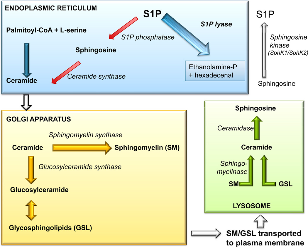
As the final enzyme in the sphingolipid degradative pathway, SPL commands the only exit point for sphingolipid intermediates and their flow into phospholipid metabolism. So, in addition to regulating S1P levels, SPL is the gatekeeper of a critical node of lipid metabolic flow. The recent crystallization of a prokaryotic SPL has provided insight into the function and potential regulation and drug targeting of this enzyme. Considering the many physiological and pathological functions of S1P signaling, it seems likely that targeting SPL to modulate S1P signaling could be useful in a variety of clinical contexts.

In this review we discuss the recent highlights related to SPL-mediated biology, the structure of the SPL protein, the function of its products, new insights regarding the usefulness of SPL targeting in treating human diseases and the consequences of permanent SPL disruption in mice.


Figures

Figure 1
Figure 1. SPL enzymatic reaction
SPL is an integral membrane protein of the ER whose enzymatic activity catalyzes the cleavage of S1P at the C2-C3 carbon bond, resulting in depletion of S1P and formation of ethanolamine phosphate and hexadecenal. The enzyme requires pyridoxal 5’-phosphate as a cofactor.
Figure 2
Figure 2. Sphingolipid metabolic pathway
The de novo biosynthesis of sphingolipids initiates in the endoplasmic reticulum, where L-serine and palmitoyl-CoA are condensed to form the initial sphingoid base, which subsequently undergoes reduction, N-acylation and desaturation to form ceramide. The newly synthesized ceramide is then transported to the golgi apparatus where it is converted to higher order sphingolipids such as sphingomyelin (SM) and glycosphingolipids (GSL). These are transported to the plasma membrane where they are used as structural components. SM and GSL can be degraded in the plasma membrane or lysosome to form ceramide which can be further deacylated back to sphingosine. Sphingosine can be phosphorylated to form sphingosine-1-phosphate (S1P). S1P can be irreversibly degraded by S1P lyase or be dephosphorylated by phosphatases and utilized for the salvage pathway (red arrows).
Figure 3
Figure 3. SPL deficiency in innate and adaptive immunity
1) SPL inhibition impairs neutrophil migration. 2) Phagocytosis of apoptotic neutrophils by macrophages and dendritic cells. 3) Increased IL-23 expression leads to increased numbers of Th-17 cells. 4) Increase in Th17 cells stimulates IL-17 secretion, which in turns increases G-CSF production. 5) G-CSF stimulates granulopoiesis, consequently resulting in neutrophilia.
Figure 4
Figure 4. Sphingoid base phosphates induce neuronal apoptosis
1) CIMES (cis-4-methylsphingosine) is taken up by the cell and phosphorylated by SphK2. 2) S1P generated in the ER activates the release of calcium from inositol 1,4,5-triphosphate-sensitive calcium pools. 3) Calpain activity is triggered by increased calcium levels and activates stress-specific caspases, causing neuronal cell death. 4) CDK5 activation and tau hyperphosphorylation found in association with sphingoid base phosphate accumulation may contribute to neuronal cell death.
Figure 5
Figure 5. Potential benefits and risks of modulating SPL for therapeutic purposes
SPL inhibition is proposed as a strategy for raising S1P levels to inhibit lymphocyte trafficking and to promote cell survival in conditions causing tissue injury. Aldehyde depletion could be of potential benefit in Sjögren–Larsson syndrome. Potential risks include promotion of carcinogenesis, inhibition of innate immune functions, perturbation of lipid homeostasis, increased acute phase reactants. SPL delivery or upregulation is proposed as a strategy for depleting S1P in conditions such as cancer, fibrosis and pathologic angiogenesis, wherein monoclonal antibodies to S1P and sphingosine kinase inhibition have shown utility. Potential risks include sensitizing non-malignant cells to cytotoxic therapy and SPL product accumulation. Key determinants of safety and efficacy will likely include the degree of SPL inhibition or activation, the timing and duration of the intervention, and tissue specificity (as determined by the bioavailability and biodistribution of SPL modulating agents).

References

    1. Alexander S, Alexander H. Lead genetic studies in Dictyostelium discoideum and translational studies in human cells demonstrate that sphingolipids are key regulators of sensitivity to cisplatin and other anticancer drugs. Semin Cell Dev Biol. 2011;22:97–104. - PubMed
    1. Allende M, Bektas M, Lee B, Bonifacino E, Kang J, Tuymetova G, et al. Sphingosine-1-phosphate lyase deficiency produces a pro-inflammatory response while impairing neutrophil trafficking. J Biol Chem. 2010;286:7348–7358. - PMC - PubMed
    1. Alvarez SE, Harikumar KB, Hait NC, Allegood J, Strub GM, Kim EY, et al. Sphingosine-1-phosphate is a missing cofactor for the E3 ubiquitin ligase TRAF2. Nature. 2010;465:1084–1088. - PMC - PubMed
    1. Bagdanoff J, Donoviel M, Nouraldeen A, Carlsen M, Jessop T, Tarver J, et al. Inhibition of sphingosine 1-Phosphate lyase for the treatment of rheumatoid arthritis: discovery of (E)-1-(4-((1R,2S,3R)-1,2,3,4-tetrahydroxybutyl)-1H-imidazol-2-yl)ethanone Oxime (LX2931) and (1R,2S,3R)-1-(2-(Isoxazol-3-yl)-1H-imidazol-4-yl)butane-1,2,3,4-tetraol (LX2932) J Med Chem. 2010;53:8650–8652. - PubMed
    1. Bandhuvula P, Honbo N, Wang GY, Jin ZQ, Fyrst H, Zhang M, et al. S1P lyase: a novel therapeutic target for ischemia/reperfusion injury of the heart. Am J Physiol Heart Circ Physiol. 2011 H1753–H61. - PMC - PubMed

Publication types

Substances