. 2011 Oct;121(10):4043-55.
doi: 10.1172/JCI57147. Epub 2011 Sep 26.

Detection of human cytomegalovirus in medulloblastomas reveals a potential therapeutic target

Affiliations

Detection of human cytomegalovirus in medulloblastomas reveals a potential therapeutic target

Ninib Baryawno et al. J Clin Invest. 2011 Oct.

Abstract

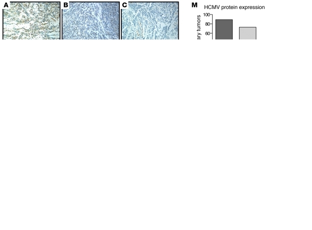
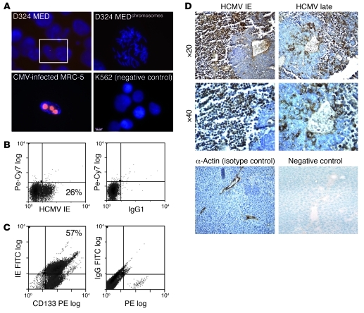
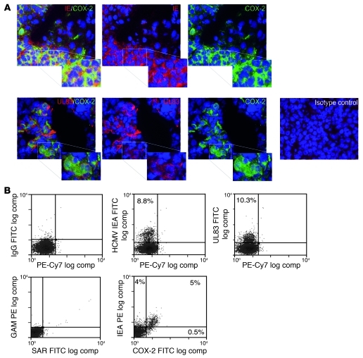
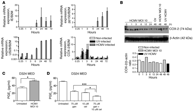
Medulloblastomas are the most common malignant brain tumors in children. They express high levels of COX-2 and produce PGE2, which stimulates tumor cell proliferation. Human cytomegalovirus (HCMV) is prevalent in the human population and encodes proteins that provide immune evasion strategies and promote oncogenic transformation and oncomodulation. In particular, HCMV induces COX-2 expression; STAT3 phosphorylation; production of PGE2, vascular endothelial growth factor, and IL-6; and tumor formation in vivo. Here, we show that a large proportion of primary medulloblastomas and medulloblastoma cell lines are infected with HCMV and that COX-2 expression, along with PGE2 levels, in tumors is directly modulated by the virus. Our analysis indicated that both HCMV immediate-early proteins and late proteins are expressed in the majority of primary medulloblastomas. Remarkably, all of the human medulloblastoma cell lines that we analyzed contained HCMV DNA and RNA and expressed HCMV proteins at various levels in vitro. When engrafted into immunocompromised mice, human medulloblastoma cells induced expression of HCMV proteins. HCMV and COX-2 expression correlated in primary tumors, cell lines, and medulloblastoma xenografts. The antiviral drug valganciclovir and the specific COX-2 inhibitor celecoxib prevented HCMV replication in vitro and inhibited PGE2 production and reduced medulloblastoma tumor cell growth both in vitro and in vivo. Ganciclovir did not affect the growth of HCMV-negative tumor cell lines. These findings imply an important role for HCMV in medulloblastoma and suggest HCMV as a novel therapeutic target for this tumor.


Figures

Figure 1
Figure 1. Detection of HCMV DNA and proteins in a majority of primary medulloblastoma tissue samples.
(AF) Immunohistochemical assessment of HCMV IE (A and D) and late (B and E) proteins in primary medulloblastomas; α-actin served as an isotype control antibody (C and F). Original magnification, ×20 (AC) and ×40 (DF). (GI) Confirmation of immunohistochemical findings by in situ hybridization. (G) HCMV DNA. (H) Negative control. (I) Positive control (Alu). Original magnification, ×40, inset, ×100 (G); ×40 (H); ×20 (I). (JL) Indirect immunofluorescence staining for HCMV IE (J) and late (K) antigens in frozen sections of a primary medulloblastoma, and isotype control for immunofluorescence (L). Original magnification, ×20 (JL). (M) Quantification of HCMV expression in medulloblastomas positive for HCMV IE and late proteins. (N) HCMV expression graded 1–4 according to the percentage of HCMV-positive cells in 37 primary medulloblastoma tissue samples (see Methods for details). L, late. (O) Flow cytometric analysis of HCMV UL83 expression in two primary medulloblastomas (M2 and M3). Isotype antibodies served as a negative control.
Figure 2
Figure 2. Detection of HCMV DNA and proteins in medulloblastoma cell lines and human medulloblastoma xenografts.
(A) FISH analysis showing the presence of HCMV DNA (red) in colcemid-treated D324 MED cells and HCMV-infected (AD169) MRC-5 cells in metaphase, but not in uninfected K562 cells. (B) Flow cytometric analysis of D324 MED cells stained for the HCMV IE antigen or an isotype control antibody (IgG1). (C) Flow cytometric analysis of D283 MED cells double-stained for HCMV IE and CD133. (D) Detection of HCMV IE and late proteins in established human medulloblastoma xenografts in NMRI nu/nu mice. α-Actin served as an isotype control.
Figure 3
Figure 3. COX-2 expression in primary medulloblastomas and cell lines is restricted to positivity for HCMV proteins.
The HCMV protein UL83 and COX-2 are coexpressed in primary medulloblastomas, as shown by (A) immunohistochemical double staining for COX-2 (green) and either HCMV IE1/IE2 protein (red) or HCMV protein UL83 (red). Nuclei were stained with DAPI (blue). Isotype antibodies served as negative control. Original magnification, ×40. (B) Flow cytometric analysis of UW228-3 cells. Top row: Cells were stained for isotype control (IgG1), IE antigen (IEA), or UL83. Bottom row: Cells were stained with the secondary antibodies goat anti-mouse (GAM) and swine anti-rabbit (SAR) (negative control) or for IE antigen and COX-2.
Figure 4
Figure 4. HCMV induces COX-2 expression and PGE2 synthesis in medulloblastoma cells.
(A) HCMV IE mRNA, US28 mRNA, and low levels of the late gene pp150 mRNA were expressed in D324 MED cells after in vitro infection with HCMV VR1814 as shown by quantitative PCR at 15 minutes to 72 hours after infection. COX-2 mRNA was detected when US28 was expressed, but earlier than IE mRNA. (BD) Superinfection of HCMV VR1814 increases COX-2 expression and PGE2 levels in D324 MED cells as shown by Western blotting (B). ctr, control. (C) PGE2 levels measured by ELISA 48 hours after infection with HCMV VR1814. (D) Ganciclovir (gan) and celecoxib (cel) significantly inhibit PGE2 synthesis in D324 MED cells as measured by PGE2 ELISA. *P < 0.05, ***P < 0.0001 (1-way ANOVA). Values are mean ± SD.
Figure 5
Figure 5. Ganciclovir and celecoxib significantly impair the growth of medulloblastoma cell lines in vitro.
(A) Ganciclovir (gan) significantly inhibits the clonogenic capacity of D324 MED, D283 MED, and UW228-3 cells in a dose-dependent manner but has no growth-inhibiting effect on HCMV-negative PC-3 or BxPC-3 cells. ***P < 0.0001 (1-way ANOVA). (B) Ganciclovir significantly augments the growth-inhibitory effect of celecoxib in D324 MED, UW228-3, and D283 MED cells. At the same concentrations that significantly inhibited the growth of medulloblastoma cells, neither drug alone nor the combinations of the two drugs affected the survival of MRC-5 cells in survival assays (MTT) in human MRC-5 fibroblasts. Values are mean ± SD.
Figure 6
Figure 6. Antiviral treatment reduces CMV protein expression and clonogenic capacity of medulloblastoma cells in vitro.
(A) D283 MED cells pretreated with 75 μM ganciclovir (D283 MEDgan) exhibited markedly reduced HCMV IE and pp65 protein expression (left panel), had significantly reduced clonogenic capacity (*P < 0.05, t test; upper-right panel, and were resistant to ganciclovir (0–150 μM) or celecoxib treatment but demonstrated reduced clonogenic capacity in treatment with a combination of ganciclovir and celecoxib (right lower panel). (B) Ganciclovir and celecoxib also inhibited the clonogenic capacity of D324 MED, D283 MED, and UW228-3 cells superinfected with HCMV VR1814. *P < 0.05, ***P < 0.0001 (1-way ANOVA). Values are mean ± SD.
Figure 7
Figure 7. Ganciclovir and celecoxib significantly impair the growth of established human medulloblastoma xenografts in NMRI nu/nu mice.
(A) Pharmacokinetics of valganciclovir (val) (plasma concentration versus time) after 1 oral dose (100 mg/kg). Solid line represents concentration-time data estimated by pharmacokinetic modeling. NMRI nu/nu mice were engrafted with 7 × 10⁶ D283 MED cells subcutaneously and randomized to receive 14 mg/kg valganciclovir twice daily, 90 mg/kg celecoxib daily, both drugs, or no treatment. All treatments were given orally through a gastric feeding tube for 12 days, starting when tumors were 0.1 ml (mean, 0.13 ml). (B and C) Valganciclovir augments the inhibitory effect of celecoxib on medulloblastoma growth in vivo, as shown by the TVI (B) and tumor weight (C) at autopsy. TVI values are mean ± SD; tumor weight is shown as median and quartiles (TVI, 2-tailed ANOVA; tumor weight, 1-way ANOVA). (D and E) Clusters of cells positive for HCMV late protein in xenograft tissues were reduced by 80% by valganciclovir and celecoxib treatment. Untreated (n = 5), 75 ± 30; valganciclovir + celecoxib (n = 7), 15 ± 5. Values are mean ± SD. ***P < 0.0001 (t test). NMRI nu/nu mice were also engrafted subcutaneously with the HCMV-negative cell lines PC-3 (30 × 106 cells) and BxPC-3 (5 × 10⁶ cells) and randomized to receive 14 mg/kg valganciclovir by gastric feeding twice daily or no treatment for 23 days (PC-3) or 32 days (BxPC-3). (F and G) PC-3 or BxPC-3 xenograft growth was not inhibited in mice treated with ganciclovir, as shown by the TVI (F) and tumor weight (G) at autopsy. TVI values are mean ± SD; tumor weight is shown as median.

Comment in

References

    1. Thompson MC, et al. Genomics identifies medulloblastoma subgroups that are enriched for specific genetic alterations. J Clin Oncol. 2006;24(12):1924–1931. - PubMed
    1. Mulhern RK, Merchant TE, Gajjar A, Reddick WE, Kun LE. Late neurocognitive sequelae in survivors of brain tumours in childhood. Lancet Oncol. 2004;5(7):399–408. doi: 10.1016/S1470-2045(04)01507-4. - DOI - PubMed
    1. Rood BR, Macdonald TJ, Packer RJ. Current treatment of medulloblastoma: recent advances and future challenges. Semin Oncol. 2004;31(5):666–675. doi: 10.1053/j.seminoncol.2004.07.009. - DOI - PubMed
    1. Parsons DW, et al. The genetic landscape of the childhood cancer medulloblastoma. Science. 2011;331(6016):435–439. - PMC - PubMed
    1. Cobbs CS, et al. Human cytomegalovirus infection and expression in human malignant glioma. Cancer Res. 2002;62(12):3347–3350. - PubMed

Publication types

MeSH terms