. 2011 Dec;232(2):280-9.
doi: 10.1016/j.expneurol.2011.09.018. Epub 2011 Sep 17.

A mouse model of blast-induced mild traumatic brain injury

Affiliations

A mouse model of blast-induced mild traumatic brain injury

Vardit Rubovitch et al. Exp Neurol. 2011 Dec.

Abstract

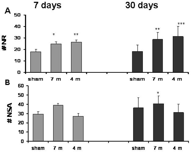
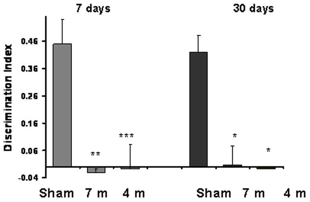
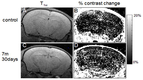
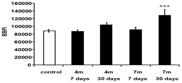
Improvised explosive devices (IEDs) are one of the main causes for casualties among civilians and military personnel in the present war against terror. Mild traumatic brain injury from IEDs induces various degrees of cognitive, emotional and behavioral disturbances but knowledge of the exact brain pathophysiology following exposure to blast is poorly understood. The study was aimed at establishing a murine model for a mild BI-TBI that isolates low-level blast pressure effects to the brain without systemic injuries. An open-field explosives detonation was used to replicate, as closely as possible, low-level blast trauma in the battlefield or at a terror-attack site. No alterations in basic neurological assessment or brain gross pathology were found acutely in the blast-exposed mice. At 7 days post blast, cognitive and behavioral tests revealed significantly decreased performance at both 4 and 7 m distance from the blast (5.5 and 2.5 PSI, respectively). At 30 days post-blast, clear differences were found in animals at both distances in the object recognition test, and in the 7 m group in the Y maze test. Using MRI, T1 weighted images showed an increased BBB permeability 1 month post-blast. DTI analysis showed an increase in fractional anisotropy (FA) and a decrease in radial diffusivity. These changes correlated with sites of up-regulation of manganese superoxide dismutase 2 in neurons and CXC-motif chemokine receptor 3 around blood vessels in fiber tracts. These results may represent brain axonal and myelin abnormalities. Cellular and biochemical studies are underway in order to further correlate the blast-induced cognitive and behavioral changes and to identify possible underlying mechanisms that may help develop treatment- and neuroprotective modalities.


Figures

Fig. 1
Fig. 1. Blast experiment setting
(a) The anesthetized mice were placed in a loose restraint device on a platform, which was covered with white plastic mesh. Each platform had space for 12 anesthetized mice. (b) Photograph of the explosive charge (A) and mice immediately prior to detonation. A cast of 500g TNT (A) was placed on a pedestal 1 meter above the ground. The 1 meter high platforms constraining the anesthetized mice were situated 4 meters (B) and 7 meters (C) meters from the TNT charge). Two pressure gauges were mounted at the ends of each platform (D).
Fig. 2
Fig. 2. Staircase test- the effect of blast on the number of rearing events (NR) and steps ascended (NSA) in a 3-min period
A. Rearing events (NR): Significant elevation NR was found in blasted mice both after 7 days (24.8±1.73 for 7m and 26.4±1.96 for 4m, compared with 18±1.83 in sham mice) and 30 days (28.8±6 for 7 m and 31.2±8.66 for 4 m, compared to 18.2±5.5 in sham mice). B. Steps ascended (NSA): The difference in the number of NSA reached statistical significance only in the 7 m group 7 days post blast (39±1.9 compared with 29.6±2.44 in sham mice). *p<0.05, **p<0.01 or ***p<0.001.
Fig. 3
Fig. 3. The effect of blast on visual memory as assessed by the novel object recognition test
The preference for novel objects was significantly reduced in all the blast groups both at 7 days(−0.02±0.01 for 7 m group, −0.008±0.009 for 4 m group and 0.45±0.09 for sham group) and 30 days (0.007±0.07 for 7 m group, −0.008±0.01 for 4 m group and 0.42±0.06 for sham group). *p<0.05,**p<0.01 or ***p<0.001.
Fig. 4
Fig. 4. The effect of blast on spatial memory as assessed by the Y-maze test
Preference for the new arm was significantly reduced in mice 7 days post blast in both groups (0.2±0.07 for 7 m group and 0.22±0.07 for 4 m in comparison with the sham group 0.51±0.15). Similar impaired memory was found after 30 days for the 4 m group (0.033±0.01 for the 4 m group and 0.361±0.09 for the sham group). *p<0.05,**p<0.01 or ***p<0.001.
Fig 5
Fig 5. BBB permeability
Contrast enhanced MRI and % change of contrast of a control mouse (A, B) and 7m 30 days post-blast mouse (D, E). The scale shows the % change of contrast enhancement 9 min post i.p. Gd-DTPA injection. There were more bright points in the brain region in E as compared to B (approximately Bregma −2.4 mm). (C, F) Respective T2w anatomical images of the same slices (TR/TE 3000/80).
Fig. 6
Fig. 6. Blast effect on BBB permeability index (BBPi)
Permeability increased after blast-explosion at 30 days at 7 m compared with control group (F(4,40)=5.9, P<0.001). *** indicates significant difference between the 7m 30 days post-blast group compared to the control group, p<0.001.
Figure 7
Figure 7. Voxel-wise statistical analysis of the whole brain DTI
Representative statistical maps overlaid on the fractional anisotropy (FA) (A) and radial diffusivity (λ3) (B) (approximately Bregma −1.3 mm). The color coded clusters show regions that present significant differences between control and blast-exposed mice (p<0.05, FDR corrected). (C-E) Quantitative values at the indicated regions of interest for control and blast-exposed mice groups of FA (C&D) at the hypothalamus and thalamus, respectively and λ3 (E) at the hypothalamus. Significant difference between the blast group and the control group *p<0.05, **p<0.01, ***p<0.001(one-way ANOVA). Control n=19, 4m 7 days n=12, 4m 7d n=7, 4m 30d n=8 and 7m 30d n=12.
Figure 8
Figure 8. FA statistical maps overlaid on the fractional anisotropy (FA) maps
Apparent regions of FA abnormality are diffusely spread over most of the brain including the cortex and the thalamus (approximately Bregma 1 mm, −1.4 mm and −2.3 mm - from left to right A-C). One-way ANOVA with significance of p<0.01 uncorrected.
Figure 9
Figure 9. Histopathology and distribution of MnSOD2 in the interpeduncular region and mammillary body after blast exposure
The left column shows low magnification photomicrographs of horizontal sections through the hypothalamus and interpeduncular nucleus (IP) of mice exposed 72h earlier to a single 5.5 PSI overpressure blast. The left upper panel is stained with hematoxylin and eosin and shows normal vasculature and brain parenchyma. The lower left panels are stained immunohistochemically for MnSOD2. Note the strong up-regulation of MnSOD2 in the mammillary body (Mm) after blast exposure, shown at higher magnification in the right panels. The calibration bar is 500 microns for the left panels and 50 microns for the right panels. Abbreviations: IP-interpeduncular nucleus, CC-crus cerebri, Mm-mammillary body, f-fornix.
Figure 10
Figure 10
Expression of C-X-C motif chemokine receptor 3 in fiber tracts after blast exposure. Photomicrographs show the emergence of immunoreactivity in association with blood vessels within 72h of a single low level blast exposure in the crus cerebri (upper row), fornix (middle row) and optic tract (lower row). The positive control staining of bone marrow from the same blast exposed and control sections are shown in the insert. The calibration bar represents 100 microns.

References

    1. Aggleton JP, Keen S, Warburton EC, Bussey TJ. Extensive cytotoxic lesions involving both the rhinal cortices and area TE impair recognition but spare spatial alternation in the rat. Brain Research Bulletin. 1997;43:279–287. - PubMed
    1. Andreasen NC. Acute and delayed posttraumatic stress disorders: a history and some issues. The American Journal of Psychiatry. 2004;161:1321–1323. - PubMed
    1. Arciniegas D, et al. Attention and memory dysfunction after traumatic brain injury: cholinergic mechanisms, sensory gating, and a hypothesis for further investigation. Brain Inj. 1999;13:1–13. - PubMed
    1. Barnum CJ, Tansey MG. (in press) The duality of TNF signaling outcomes in the brain: Potential mechanisms? Experimental Neurology. 2011 (in press) - PubMed
    1. Basser PJ, Pierpaoli C. Microstructural and physiological features of tissues elucidated by quantitative-diffusion-tensor MRI. Journal of Magnetic Resonance. 1996;111:209–219. - PubMed

Publication types

MeSH terms