. 2011 Sep 25;7(11):818-26.
doi: 10.1038/nchembio.670.

Affinity-based proteomics reveal cancer-specific networks coordinated by Hsp90

Affiliations

Affinity-based proteomics reveal cancer-specific networks coordinated by Hsp90

Kamalika Moulick et al. Nat Chem Biol. .

Abstract

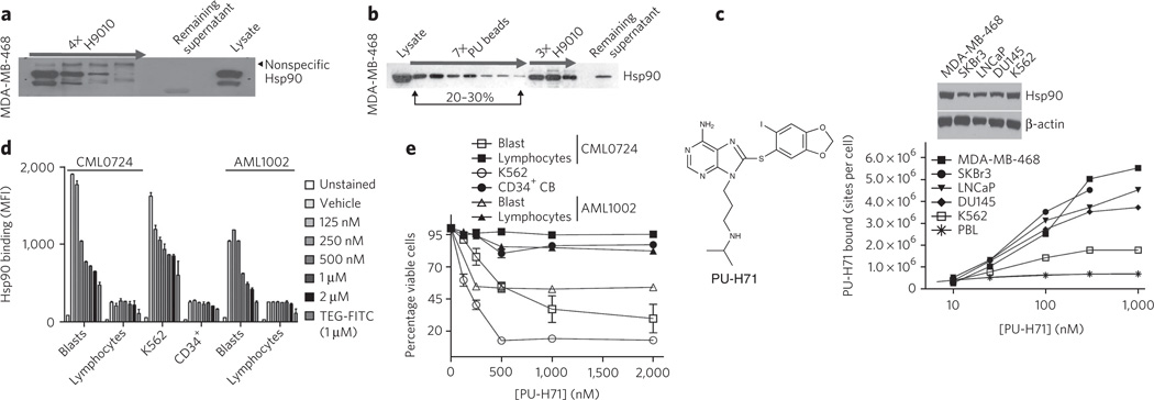
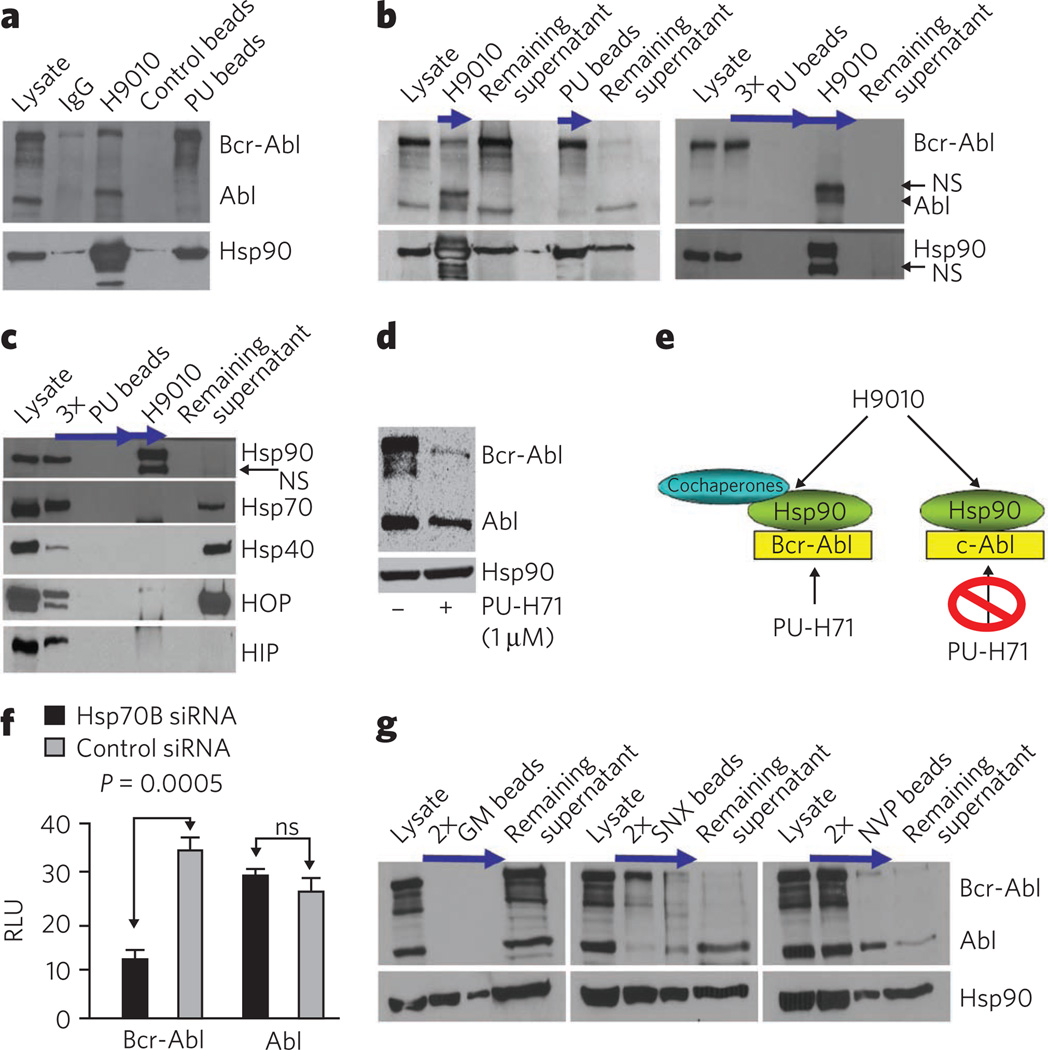
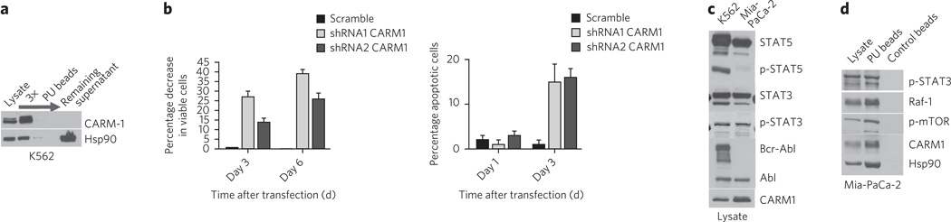
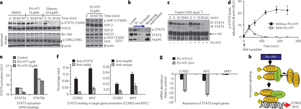
Most cancers are characterized by multiple molecular alterations, but identification of the key proteins involved in these signaling pathways is currently beyond reach. We show that the inhibitor PU-H71 preferentially targets tumor-enriched Hsp90 complexes and affinity captures Hsp90-dependent oncogenic client proteins. We have used PU-H71 affinity capture to design a proteomic approach that, when combined with bioinformatic pathway analysis, identifies dysregulated signaling networks and key oncoproteins in chronic myeloid leukemia. The identified interactome overlaps with the well-characterized altered proteome in this cancer, indicating that this method can provide global insights into the biology of individual tumors, including primary patient specimens. In addition, we show that this approach can be used to identify previously uncharacterized oncoproteins and mechanisms, potentially leading to new targeted therapies. We further show that the abundance of the PU-H71-enriched Hsp90 species, which is not dictated by Hsp90 expression alone, is predictive of the cell's sensitivity to Hsp90 inhibition.


Figures

Figure 1
Figure 1. PU-H71 interacts with a restricted fraction of Hsp90 that is more abundant in cancer cells
(a) Sequential immunopurification steps, as indicated by the arrow, with H9010 (a Hsp90-specific antibody) deplete Hsp90 in the MDA-MB-468 cell extract. Lysate, control cell extract. (b) Hsp90 from MDA-MB-468 extracts was isolated through sequential chemical-purification and immunopurification steps. (c) Saturation studies were performed with 131I-PU-H71 in the indicated cells (below). Expression of Hsp90 in the indicated cells was analyzed by western blotting (above). Representative data of four separate repeats are presented. (d) Binding of PU-FITC, presented as mean fluorescence intensity (MFI), to primary AML and CML, CD34+ cord blood cells (CB), or K562 cells pretreated with the indicated doses of PU-H71 for 24 h. TEG-FITC is a nonspecific binding control. (e) Percent viability relative to untreated control for the indicated cells after treatment for 96 h with the indicated doses of PU-H71. Data are presented as means ± s.e.m. (n = 3).
Figure 2
Figure 2. PU-H71 is selective for and isolates Hsp90 in complex with oncoproteins and cochaperones
(a) Representative western blot of Hsp90 complexes in K562 extracts isolated by precipitation with H9010, a nonspecific IgG, PU beads or control beads containing 2-methoxyethylamine, an Hsp90-inert molecule. (b,c) Single or sequential immunoprecipitations and chemical precipitations, as indicated by arrows, conducted in K562 extracts with H9010 and PU beads. NS, nonspecific. (d) Representative western blot of K562 cells treated for 24 h with vehicle (−) or PU-H71 (+). (e) Proposed Hsp90 species in K562 cells, in complex with both aberrant, Bcr-Abl, and normal, c-Abl, proteins. PU-H71, but not H9010, selects for the Hsp90 population that is Bcr-Abl oncoprotein bound. (f) Expression of proteins in Hsp70–knocked down K562 cells. Changes in protein concentrations are presented in relative luminescence units (RLU). Control, scrambled small interfering RNA (siRNA). Data are presented as means ± s.e.m. (n = 3). ns, nonsignificant. (g) Representative western blot of sequential chemical precipitations, as indicated by arrows, conducted in K562 extracts with GM, SNX and NVP beads.
Figure 3
Figure 3. PU-H71 identifies the aberrant signalosome in CML cells
(a) Pathway diagram highlighting the PU bead–identified CML signalosome with focus on networks 1 (Raf-MAPK and PI3K-AKT-mTO R pathway), 2 (NF-κB pathway) and 8 (STAT5 pathway). Key nodal proteins in the identified networks are shown in yellow. A detailed list of identified protein networks and component proteins is shown in Supplementary Figure 6 and Supplementary Data Set 1f. (b) Left, representative western blot of a subset of MS-identified protein complexes. No proteins were detected in the control bead pull-downs, and those data are omitted for simplicity of presentation. Right, representative western blot of K562 cells treated for 24 h with vehicle (−) or PU-H71 (+). (c) Representative western blot of single chemical precipitations conducted in primary CML cell extracts with PU and control beads.
Figure 4
Figure 4. PU-H71-identified proteins and networks are those important for the malignant phenotype
(a) Representative western blot of sequential chemical precipitations, as indicated by the arrow, conducted in K562 extracts with the PU beads. (b) The effect of CARM1 knockdown on cell viability using tryptan blue (left) or acridine orange-ethidium bromide (right) staining was evaluated in K562 cells. Data are presented as means ± s.e.m. (n = 3). (c) The expression of select potential Hsp90-interacting proteins was analyzed by western blotting in K562 leukemia and Mia-PaCa-2 pancreatic cancer cells. (d) Representative western blot of single chemical precipitations conducted in Mia-PaCa-2 cell extracts with PU and control beads.
Figure 5
Figure 5. Hsp90 facilitates an enhanced STAT5 activity in CML
(a) Representative western blot of K562 cells treated for the indicated times with PU-H71 (5 µM), Gleevec (0.5 µM) or DMSO (vehicle). (b) Representative western blot of sequential chemical precipitations conducted in K562 cells with PU and control beads, as indicated by the blue arrow. (c) Representative western blot of STAT5 immunocomplexes from cells pretreated with vehicle or PU-H71, and then treated for the indicated times with trypsin. (d) p-STAT5 concentrations in K562 cells treated for the indicated times with vanadate (1 mM) in the presence and absence of PU-H71 (5 µM). Data are presented as mean ± s.d. (n = 3). (e) The DNA-binding capacity of STAT5 in K562 cells treated for 24 h with the indicated concentrations of PU-H71. (f) Quantitative ChIP performed with STAT5 or Hsp90 antibodies versus an IgG control for two known STAT5 target genes. A primer that amplifies an intergenic region was used as negative control. Results are expressed as a percentage of the input for the specific antibody (STAT5 or Hsp90) over the respective IgG control. (g) The transcript abundance of CCND2 and MYC in K562 cells exposed to µM of PU-H71. Results are expressed as fold change compared to baseline (time 0 h) and were normalized to RPL13A. HPRT was used as negative control. Data are presented as means ± s.e.m. (h) Proposed mechanism for Hsp90-facilitated increased STAT5 signaling in CML. Hsp90 binds to and influences the conformation of STAT5 and maintains STAT5 in an active conformation directly within STAT5-containing transcriptional complexes.

References

    1. Ley TJ, et al. DNA sequencing of a cytogenetically normal acute myeloid leukaemia genome. Nature. 2008;456:66–72. - PMC - PubMed
    1. Parsons DW, et al. An integrated genomic analysis of human glioblastoma multiforme. Science. 2008;321:1807–1812. - PMC - PubMed
    1. Hanash S, Taguchi A. The grand challenge to decipher the cancer proteome. Nat. Rev. Cancer. 2010;10:652–660. - PubMed
    1. Kolch W, Pitt A. Functional proteomics to dissect tyrosine kinase signalling pathways in cancer. Nat. Rev. Cancer. 2010;10:618–629. - PubMed
    1. Nomura DK, Dix MM, Cravatt BF. Activity-based protein profiling for biochemical pathway discovery in cancer. Nat. Rev. Cancer. 2010;10:630–638. - PMC - PubMed

Publication types

Substances