

Quantitative assessment of artifacts on cardiac magnetic resonance imaging of patients with pacemakers and implantable cardioverter-defibrillators
- PMID: 21946701
- PMCID: PMC3218212
- DOI: 10.1161/CIRCIMAGING.111.965764
Quantitative assessment of artifacts on cardiac magnetic resonance imaging of patients with pacemakers and implantable cardioverter-defibrillators
Abstract
Background: The safety and clinical utility of MRI at 1.5 T in patients with cardiac implantable devices such as pacemakers (PM) and implantable cardioverter-defibrillators (ICD) have been reported. This study aims to evaluate the extent of artifacts on cardiac magnetic resonance (CMR) in patients with PM and ICD (PM/ICD).
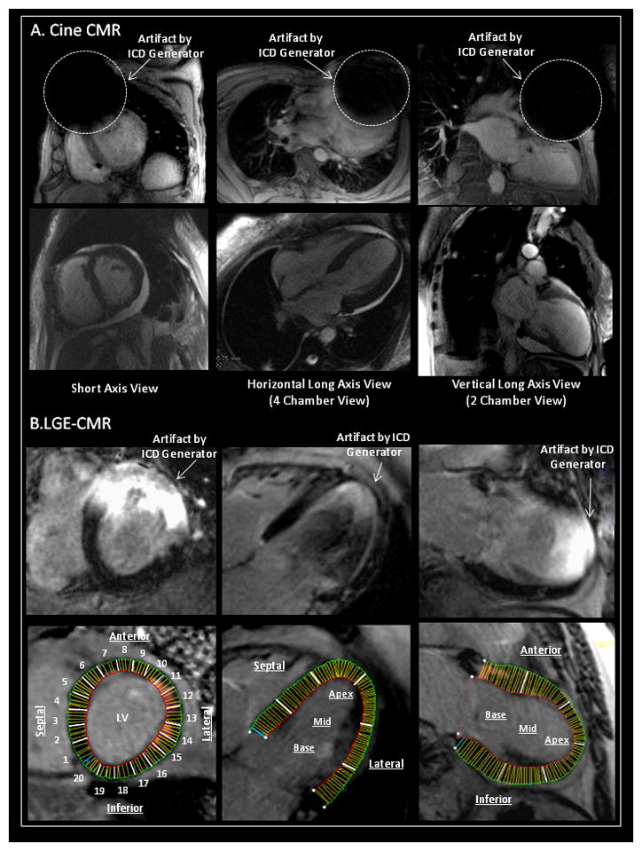
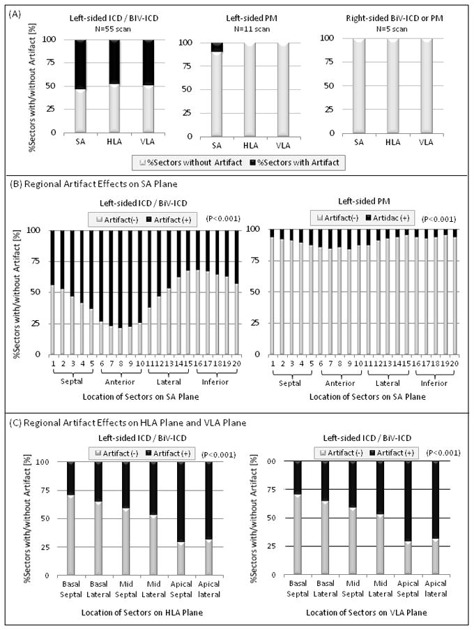
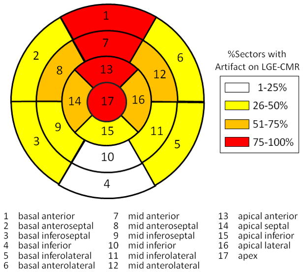
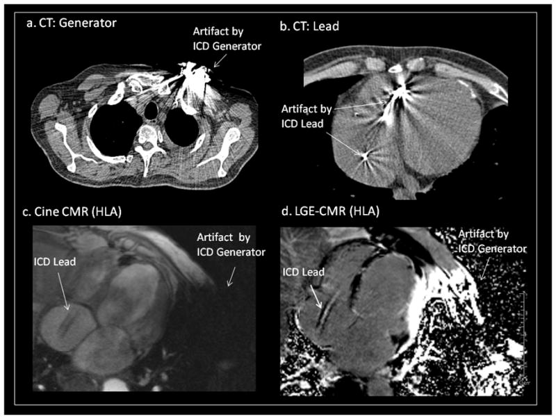
Methods and results: A total of 71 CMR studies were performed with an established safety protocol in patients with prepectoral PM/ICD. The artifact area around the PM/ICD generator was measured in all short-axis (SA), horizontal (HLA), and vertical long-axis (VLA) SSFP cine planes. The location and extent of artifacts were also assessed in all SA (20 sectors per plane), HLA, and VLA (6 sectors per plane) late gadolinium-enhanced CMR (LGE-CMR) planes. The artifact area on cine CMR was significantly larger with ICD versus PM generators in each plane (P<0.001, respectively). In patients with left-sided ICD or biventricular ICD systems, the percentages of sectors with any artifacts on LGE-CMR were 53.7%, 48.0%, and 49.2% in SA, HLA, and VLA planes, respectively. Patients with left-sided PM or right-sided PM/ICD had fewer artifacts. Anterior and apical regions were severely affected by artifact caused by left-sided PM/ICD generators.
Conclusions: In contrast to patients with right-sided PM/ICD and left-sided PM, the anterior and apical left ventricle can be affected by susceptibility artifacts in patients with left-sided ICD. Artifact reduction methodologies will be necessary to improve the performance of CMR in patients with left sided ICD systems.
Figures
References
-
- Nazarian S, Roguin A, Zviman MM, Lardo AC, Dickfeld TL, Calkins H, Weiss RG, Berger RD, Bluemke DA, Halperin HR. Clinical utility and safety of a protocol for noncardiac and cardiac magnetic resonance imaging of patients with permanent PMs and ICDs at 1.5 tesla. Circulation. 2006;114:1277–1284. - PMC - PubMed
-
- Nazarian S, Halperin HR. How to perform magnetic resonance imaging on patients with implantable cardiac arrhythmia devices. Heart Rhythm. 2009;6:138–143. - PubMed
-
- Pulver AF, Puchalski MD, Bradley DJ, Minich LL, SUJT, Saarel EV, Whitaker P, Etheridge SP. Safety and imaging quality of MRI in pediatric and adult congenital heart disease patients with PMs. PACE. 2009;32:450–456. - PubMed
-
- Naehle CP, Strach K, Thomas D, Meyer C, Linhart M, Bitaraf S, Litt H, Schwab JO, Schild H, Sommer T. Magnetic resonance imaging at 1.5-T in patients with ICDs. J Am Coll Cardiol. 2009;54:549–555. - PubMed
Publication types
MeSH terms
Grants and funding
LinkOut - more resources
Full Text Sources
Other Literature Sources
Medical
Research Materials
