

CD140a identifies a population of highly myelinogenic, migration-competent and efficiently engrafting human oligodendrocyte progenitor cells
- PMID: 21947029
- PMCID: PMC3365580
- DOI: 10.1038/nbt.1972
CD140a identifies a population of highly myelinogenic, migration-competent and efficiently engrafting human oligodendrocyte progenitor cells
Abstract
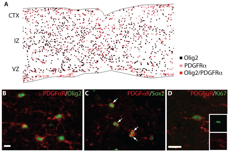
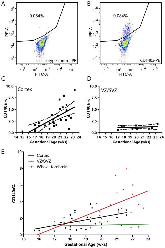
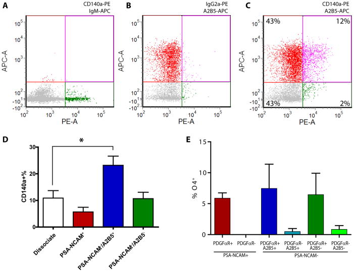
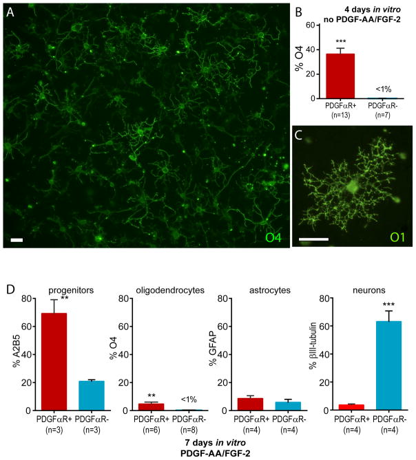
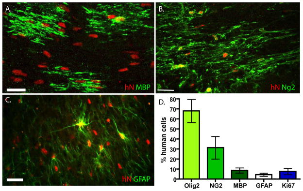
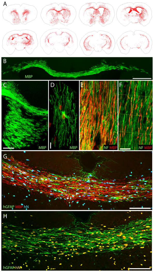
Experimental animals with myelin disorders can be treated by transplanting oligodendrocyte progenitor cells (OPCs) into the affected brain or spinal cord. OPCs have been isolated by their expression of gangliosides recognized by mAb A2B5, but this marker also identifies lineage-restricted astrocytes and immature neurons. To establish a more efficient means of isolating myelinogenic OPCs, we sorted fetal human forebrain cells for CD140a, an epitope of platelet derived growth factor receptor (PDGFR)α, which is differentially expressed by OPCs. CD140a(+) cells were isolated as mitotic bipotential progenitors that initially expressed neither mature neuronal nor astrocytic phenotypic markers, yet could be instructed to either oligodendrocyte or astrocyte fate in vitro. Transplanted CD140a(+) cells were highly migratory and robustly myelinated the hypomyelinated shiverer mouse brain more rapidly and efficiently than did A2B5(+)cells. Microarray analysis of CD140a(+) cells revealed overexpression of the oligodendroglial marker CD9, suggesting that CD9(+)/CD140a(+) cells may constitute an even more highly enriched population of myelinogenic progenitor cells.
Figures
Comment in
-
Tracking down the human myelinating cell.Nat Biotechnol. 2011 Oct 13;29(10):881-3. doi: 10.1038/nbt.2004. Nat Biotechnol. 2011. PMID: 21997628 No abstract available.
Similar articles
-
Human fetal oligodendrocyte progenitor cells from different gestational stages exhibit substantially different potential to myelinate.Stem Cells Dev. 2012 Jul 20;21(11):1831-7. doi: 10.1089/scd.2011.0494. Epub 2012 Jan 26. Stem Cells Dev. 2012. PMID: 22122665
-
Anti-muscarinic adjunct therapy accelerates functional human oligodendrocyte repair.J Neurosci. 2015 Feb 25;35(8):3676-88. doi: 10.1523/JNEUROSCI.3510-14.2015. J Neurosci. 2015. PMID: 25716865 Free PMC article.
-
Human iPSC-derived oligodendrocyte progenitor cells can myelinate and rescue a mouse model of congenital hypomyelination.Cell Stem Cell. 2013 Feb 7;12(2):252-64. doi: 10.1016/j.stem.2012.12.002. Cell Stem Cell. 2013. PMID: 23395447 Free PMC article.
-
Redox state as a central modulator of precursor cell function.Ann N Y Acad Sci. 2003 Jun;991:251-71. doi: 10.1111/j.1749-6632.2003.tb07481.x. Ann N Y Acad Sci. 2003. PMID: 12846992 Review.
-
Remyelination of the demyelinated CNS: the case for and against transplantation of central, peripheral and olfactory glia.Brain Res Bull. 2002 Apr;57(6):827-32. doi: 10.1016/s0361-9230(01)00765-1. Brain Res Bull. 2002. PMID: 12031280 Review.
Cited by
-
Optimizing culture medium composition to improve oligodendrocyte progenitor cell yields in vitro from subventricular zone-derived neural progenitor cell neurospheres.PLoS One. 2015 Apr 2;10(4):e0121774. doi: 10.1371/journal.pone.0121774. eCollection 2015. PLoS One. 2015. PMID: 25837625 Free PMC article.
-
Glial progenitor cell-based treatment of the childhood leukodystrophies.Exp Neurol. 2016 Sep;283(Pt B):476-88. doi: 10.1016/j.expneurol.2016.05.010. Epub 2016 May 8. Exp Neurol. 2016. PMID: 27170209 Free PMC article. Review.
-
Glia Disease and Repair-Remyelination.Cold Spring Harb Perspect Biol. 2015 May 18;7(7):a020594. doi: 10.1101/cshperspect.a020594. Cold Spring Harb Perspect Biol. 2015. PMID: 25986556 Free PMC article. Review.
-
Paired Related Homeobox Protein 1 Regulates Quiescence in Human Oligodendrocyte Progenitors.Cell Rep. 2018 Dec 18;25(12):3435-3450.e6. doi: 10.1016/j.celrep.2018.11.068. Cell Rep. 2018. PMID: 30566868 Free PMC article.
-
Remyelination in PNS and CNS: current and upcoming cellular and molecular strategies to treat disabling neuropathies.Mol Biol Rep. 2021 Dec;48(12):8097-8110. doi: 10.1007/s11033-021-06755-6. Epub 2021 Nov 3. Mol Biol Rep. 2021. PMID: 34731366 Review.
References
-
- Ben-Hur T, Goldman SA. Prospects of cell therapy for disorders of myelin. Ann N Y Acad Sci. 2008;1142:218–249. - PubMed
-
- Franklin RJ, Ffrench-Constant C. Remyelination in the CNS: from biology to therapy. Nat Rev Neurosci. 2008;9:839–855. - PubMed
-
- Scolding N, et al. Oligodendrocyte progenitors are present in the normal adult human CNS and in the lesions of multiple sclerosis. Brain. 1998;121:2221–2228. - PubMed
Publication types
MeSH terms
Substances
Grants and funding
LinkOut - more resources
Full Text Sources
Other Literature Sources
Medical
Molecular Biology Databases
Miscellaneous
