. 2011 Dec;31(23):4720-34.
doi: 10.1128/MCB.06147-11. Epub 2011 Sep 26.

SIRT1 deacetylates the DNA methyltransferase 1 (DNMT1) protein and alters its activities

Affiliations

SIRT1 deacetylates the DNA methyltransferase 1 (DNMT1) protein and alters its activities

Lirong Peng et al. Mol Cell Biol. 2011 Dec.

Abstract

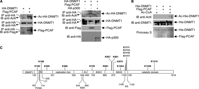
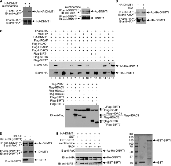
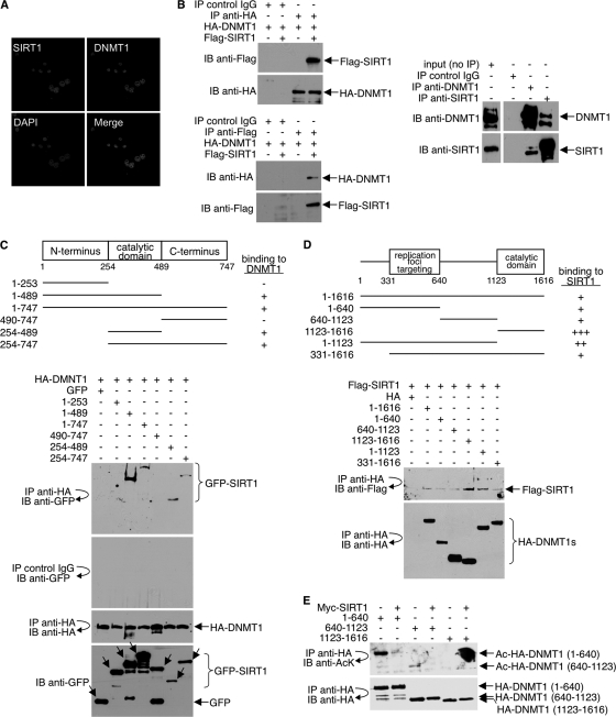
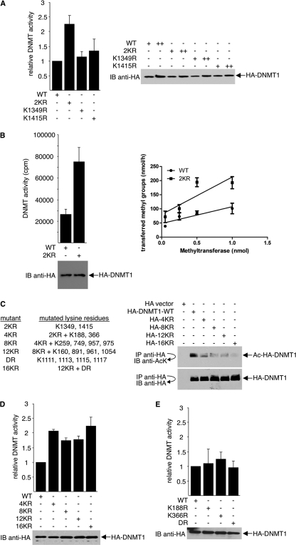
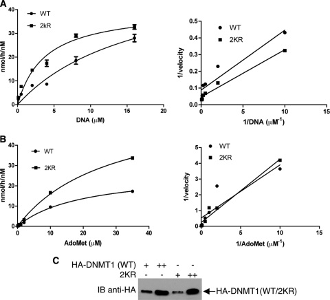
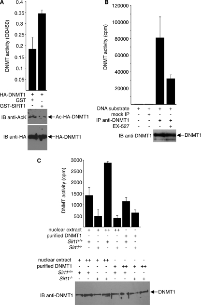
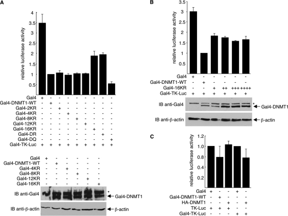
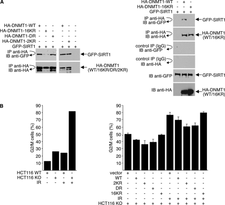
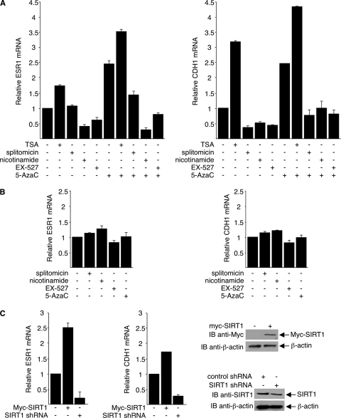
DNA methylation and histone acetylation/deacetylation are distinct biochemical processes that control gene expression. While DNA methylation is a common epigenetic signal that inhibits gene transcription, histone deacetylation similarly represses transcription but can be both an epigenetic and nonepigenetic phenomenon. Here we report that the histone deacetylase SIRT1 regulates the activities of DNMT1, a key enzyme responsible for DNA methylation. In mass spectrometry analysis, 12 new acetylated lysine sites were identified in DNMT1. SIRT1 physically associates with DNMT1 and can deacetylate acetylated DNMT1 in vitro and in vivo. Interestingly, deacetylation of different lysines on DNMT1 has different effects on the functions of DNMT1. For example, deacetylation of Lys1349 and Lys1415 in the catalytic domain of DNMT1 enhances DNMT1's methyltransferase activity, while deacetylation of lysine residues in the GK linker decreases DNMT1's methyltransferase-independent transcriptional repression function. Furthermore, deacetylation of all identified acetylated lysine sites in DNMT1 abrogates its binding to SIRT1 and impairs its capability to regulate cell cycle G(2)/M transition. Finally, inhibition of SIRT1 strengthens the silencing effects of DNMT1 on the expression of tumor suppressor genes ER-α and CDH1 in MDA-MB-231 breast cancer cells. Together, these results suggest that SIRT1-mediated deacetylation of DNMT1 is crucial for DNMT1's multiple effects in gene silencing.


Figures

Fig. 1.
Fig. 1.
Acetylation of DNMT1 in vivo and in vitro. (A) 293T cells were transfected with HA-DNMT1 and either Flag-PCAF, HA-p300, or empty Flag vector. Cell lysates were immunoprecipitated (IP) with antibody to HA, and immunoprecipitates were immunoblotted (IB) with antibody to HA, Flag, or acetyllysine (AcK). Ac, acetylated. Direct immunoblot assays were performed with anti-Flag and anti-HA to assess expressions of Flag-PCAF and HA-p300, respectively. (B) His-DNMT1 was expressed in insect cells using the baculovirus system and purified on nickel affinity columns. Flag-PCAF was expressed in 293T cells and immunopurified from cell lysates with an anti-Flag antibody. In vitro acetylation reaction mixtures contained His-DNMT1, Flag-PCAF, and acetyl-CoA as indicated. Reaction products were detected with an antiacetyllysine antibody. Anti-DNMT1 Western blots and Ponceau S staining show equal amounts of His-DNMT1 and Flag-PCAF in reaction mixtures. (C) 293T cells expressing HA-DNMT1 were treated with 15 mM nicotinamide plus 100 ng/ml TSA overnight. HA-DNMT1 was immunoprecipitated from cell lysates with antibody to HA, and immunoprecipitated material was analyzed by mass spectrometry. Acetylated lysines identified in the analysis, plus previously reported acetylation sites (Lys111, Lys1113, Lys1115, and Lys1117), are indicated in bold type. The domains of DNMT1 are shown and are drawn in approximate scale. DMAP1, DNA methyltransferase 1-associated protein 1-binding domain; PCNA, proliferating cell nuclear antigen-binding domain; N, nuclear localization signal; replication foci, replication focus-targeting domain; KEN, KEN box (KENXXXR sequence); Zn, zinc finger region; BAH1 and BAH2, bromo-adjacent homology domains; KG, lysine-glycine repeats, also called GK linker.
Fig. 2.
Fig. 2.
Deacetylation of DNMT1 by SIRT1 in vivo and in vitro. (A) Anti-HA (left) or anti-DNMT1 (right) immunoprecipitates from 293T cells, which were transfected with HA-DNMT1 expression plasmid (left panel) or untransfected (right panel) and treated with 15 mM nicotinamide for 12 h, were immunoblotted with antibody to AcK. The membranes were stripped and reprobed with anti-HA or anti-DNMT1. (B) 293T cells expressing HA-DNMT1 received 400 ng/ml of TSA for 2 h. Anti-HA immunoprecipitates were immunoblotted with antibody to acetyllysine or HA. (C) 293T cells were transfected with HA-DNMT1, Flag-PCAF, and Flag-HDAC1, -2, or -3 or Flag-SIRT1, -6, or -7 as indicated. Cell lysates were immunoprecipitated with antibody to HA or were mock precipitated (IgG), and immunoprecipitates were immunoblotted (IB) with antibody to acetyllysine. The membrane was stripped and reprobed with anti-HA. A separate Western blot assay was performed with anti-Flag antibody to assess Flag-HDAC and Flag-SIRT expressions (bottom). (D) HeLa cells were transfected with either shRNA pSuper-SIRT1 (HeLa-S) or scrambled control shRNA (HeLa-C) and grown in 1 μg/ml puromycin for 2 weeks. SIRT1 depletion in HeLa-S cells was assessed by Western blot assays, and one colony (HeLa-S5) was selected for further analysis. Anti-DNMT1 immunoprecipitates were immunoblotted with antibodies to acetyllysine or DNMT1. Immunoblot assays were also performed to assess SIRT1 expression. (E) (Left) HA-DNMT1 was first hyperacetylated in vivo by coexpression with PCAF in 293T cells, and then cell lysates were prepared and immunoprecipitated with antibody to HA. In vitro deacetylation reactions were performed by incubating anti-HA immunoprecipitates (Ac-HA-DNMT1), 1 μg GST or GST-SIRT1, NAD+, and 10 mM nicotinamide as indicated. Reaction products were immunoblotted with antibody to acetyllysine. The membrane was stripped and reprobed with anti-HA or anti-SIRT1. (Right) The quality of bacterially expressed, purified GST and GST-SIRT1 proteins was assessed by SDS-PAGE and Coomassie blue staining.
Fig. 3.
Fig. 3.
SIRT1 interacts with DNMT1. (A) To examine the colocalization of endogenous SIRT1 and DNMT1 in the nucleus of HeLa cells, immunofluorescence studies were performed as described in Materials and Methods. Mouse anti-DNMT1 and rabbit anti-SIRT1 antibodies were used. (B) (Left) 293T cells were cotransfected with HA-DNMT1 and Flag-SIRT1. Mock precipitates (IgG control) and anti-HA and anti-Flag immunoprecipitates were immunoblotted with anti-HA or anti-Flag antibody. (Right) IgG, anti-DNMT1, and anti-SIRT1 immunoprecipitates from 293T whole-cell lysates were immunoblotted with antibody to DNMT1 or SIRT1. (C) (Top) Schematic diagram of GFP-SIRT1 and GFP-SIRT1 deletion mutants (not drawn to scale). For simplicity, the GFP portion is not included in the illustration. The ability of wild-type (1–747) and each mutant SIRT1 to bind HA-DNMT1 is indicated (+ or −). (Bottom) 293T cells were cotransfected with full-length HA-DNMT1 and either full-length GFP-SIRT1 (1–747) or the GPF-SIRT1 deletion mutants; IgG and anti-HA immunoprecipitates were immunoblotted with antibody to GFP or HA (top three blots). Cell lysates were immunoblotted with antibody to GFP to show the expression levels of SIRT1 (bottom blot). (D) (Top) Schematic diagram of HA-DNMT1 and HA-DNMT1 deletion mutants (not drawn to scale). For simplicity, the HA portion is not included in the illustration. The ability of wild-type (1–1616) and each mutant DNMT1 to bind Flag-SIRT1 is indicated (+ or −). (Bottom) 293T cells were cotransfected with full-length Flag-SIRT1 and either full-length HA-DNMT1 or HA-DNMT1 deletion mutants. Anti-HA immunoprecipitates were washed exhaustively with a buffer containing 500 mM NaCl and 0.5% NP-40 and immunoblotted with antibody to Flag or HA. (E) 293T cells were cotransfected with the HA-DNMT1 deletion mutants and either Myc-SIRT1 or empty vector. Anti-HA immunoprecipitates were immunoblotted with antibody to antiacetyllysine (AcK) or HA.
Fig. 4.
Fig. 4.
Deacetylation of DNMT1 increases its methyltransferase activity. (A) K1349 and K1415 of HA-DNMT1 were mutated to arginine either individually (K1349R and K1415R) or together (2KR). Wild-type DNMT1 (WT) and the DNMT1 mutants were ectopically expressed in 293T cells. Equal amounts of cell lysates were assayed for DNA methyltransferase activity by an anti-methyl-cytosine antibody-based ELISA (left). Twenty micrograms (+) and 60 μg (++) of cell lysates were Western blotted with anti-HA antibody to ensure equal expression of the HA constructs in cells (right). (B) WT and 2KR mutant HA-DNMT1 were overexpressed in 293T cells. Cell lysates were analyzed for DNA methyltransferase activity using the isotope labeling method as described in Materials and Methods. After incubation at 37°C for 2 h, unincorporated nucleotides were removed and the incorporation of radioactivity was determined by liquid scintillation counting. Results of representative Western blot assays to compare WT and 2KR expressions are shown (bottom left). For both purified wild-type and 2KR DNMT1, four different concentrations were compared (right). (C) (Left) List of the lysine residues mutated in DNMT1. (Right) To examine protein acetylation levels, wild-type HA-DNMT1 and the indicated HA-DNMT1 mutants were expressed in 293T cells. Anti-HA immunoprecipitates were immunoblotted with antiacetyllysine antibody, stripped, and reprobed with anti-HA antibody. (D) The indicated wild type and HA-DNMT1 mutants were expressed in 293T cells, and cell lysates were assayed for methyltransferase activity (top). HA-DNMT1 expressions were determined using Western blot assays with anti-HA antibody, and representative results are shown (bottom). (E) The indicated wild type and HA-DNMT1 mutants were expressed in 293T cells, and cell lysates were assayed for methyltransferase activity (top). Protein expressions were determined using Western blot assays with anti-HA antibody, and representative results are shown (bottom).
Fig. 5.
Fig. 5.
Steady-state kinetics determination of wild type (WT) and 2KR mutant. (A) Kinetics of poly(dI-dC)·(dI-dC). Duplicate reaction mixtures contained either 10 nM WT or 2KR with 10 μM [3H]AdoMet (10 Ci/1 mmol) and various concentrations of DNA at 0.1, 0.5, 2, 4, 8, or 16 μM in 25 μl reaction buffer (50 mM Tris-HCl [pH 8.0], 1 mM EDTA, 5 mM DTT, 10% glycerol). (B) Kinetics for the substrate of AdoMet. Duplicate reactions were performed by incubating 10 nM enzyme, 6 μM poly(dI-dC)·(dI-dC), and titrated [3H]AdoMet at 0.1, 0.5, 1, 2, 10, and 35 μM in a 25-μl reaction volume. All reactions were performed for 1 h at 37°C, and the transferring of methyl groups was measured as described in Materials and Methods. The resultant velocities (nmol/h/nM) were plotted using GraphPad Prism5 software. (Left) Nonlinear regression of Michaelis-Menten kinetics of velocity versus DNA concentration; (right) corresponding double reciprocal (Lineweaver-Burk) plots of velocity versus DNA concentration. (C) Western blot assay to assess the quality and quantity of WT and 2KR DNMT1 used in the kinetics assays.
Fig. 6.
Fig. 6.
Increased methyltransferase activity of SIRT1-deacetylated DNMT1. (A) In vitro methyltransferase assays were performed with an ELISA-like assay. Reaction mixtures contained immunopurified HA-DNMT1s that were preincubated with either GST or GST-SIRT1 in the presence of NAD+. (Bottom) Immunoblot assays with anti-HA and anti-AcK were done to ensure equal HA-DNMT1 quantity in each assay and to assess deacetylation of DNMT1 by GST-SIRT1, respectively. (B) Endogenous DNMT1s immunoprecipitated from 293T cells, treated or untreated with EX-527, were assayed for methyltransferase activity. Negative controls include mock precipitates with IgG or without DNA substrate. Western blot assays with anti-DNMT1 were performed to ensure equal DNMT1 in each assay (bottom). (C) Nuclear extracts or immunopurified endogenous DNMT1s, prepared from Sirt1+/+ or Sirt1−/− MEFs, were assayed for methyltransferase activity. Western blot assays with anti-DNMT1 were performed to ensure equal DNMT1 in each assay (bottom). Representative blot results are shown.
Fig. 7.
Fig. 7.
Deacetylation of DNMT1 GK linker reduces DNMT1 methyltransferase-independent transcription repression activity. (A and C) 293T cells in 12-well plates were transfected with either pTK-Luc or pGal4-TK-Luc (0.1 μg), pRL-SV40 (internal control, 0.018 μg), and the indicated HA-DNMT1, Gal4, or Gal4-DNMT1 constructs (0.5 μg). (B) 293T cells in 12-well plates were transfected with pGal4-TK-Luc (0.1 μg), pRL-SV40 (internal control, 0.018 μg), and the indicated Gal4-DNMT1-WT (0.5 μg) or Gal4-16KR (0.5, 1, 1.5, and 2 μg). Firefly luciferase activity is normalized to Renilla luciferase activity and depicted as fold over wild-type value with error bars showing standard deviations from three experiments. Anti-Gal4 and anti-β-actin immunoblot assays were performed to assess Gal4-DNMT1 expression and to serve as loading controls. *, nonspecific bands on Western blots.
Fig. 8.
Fig. 8.
Deacetylation of DNMT1 affects its cell cycle regulatory function. (A) 293T cells were transfected with GFP-SIRT1 and either wild-type or mutant HA-DNMT1 expression plasmids. Cell lysates were immunoprecipitated with antibody to HA, and precipitated material was immunoblotted with antibodies to GFP or HA. GFP-SIRT1 and HA-DNMT1 expressions were monitored by direct Western blotting with anti-GFP and anti-HA antibodies, respectively. (B) Wild-type HCT116 (WT) cells or DNMT1-knockout (KO) HCT116 cells were cotransfected with GFP and either wild-type DNMT1 (WT), mutant HA-DNMT1, or vector plasmid. The ratio of GFP to HA-DNMT1 was 1:10. Cells received 10 Gy of gamma irradiation (IR) 36 h after transfection and were harvested at 48 h posttransfection. Cell cycle position was determined by FACS analysis of propidium iodide-stained cells for GFP-positive cells. The data shown are the average values ± standard deviations from three separate experiments (right).
Fig. 9.
Fig. 9.
SIRT1 regulates DNMT1-mediated silencing of TSGs. (A and B) MDA-MB-231 (A) and MCF-7 (B) cells were treated with 0.3 μM TSA for 12 h, 15 mM nicotinamide for 12 h, 300 μM splitomicin for 24 h, or 1 μM EX-527 for 6 h. Some cultures also received 50 μM 5-aza-dC for 48 h. (C) MDA-MB-231 cells with plasmids that express either Myc-SIRT1 or SIRT1 shRNA. Expressions of ESR1 and CDH1 mRNA were determined by quantitative real-time PCR. The RNA of untreated MCF-7 cells was used as positive control and for generation of a standard curve. 18S RNA was used as the internal control. Amounts of PCR-amplified ESR1 mRNA and CDH1 mRNA were determined from the standard curve and normalized to the amount of 18S RNA. Results from averages of three experiments with standard deviations are depicted as fold of untreated control. SIRT1 protein expressions were assessed using Western blot assays (right).

References

    1. Adams R. L., Rinaldi A., Seivwright C. 1991. Microassay for DNA methyltransferase. J. Biochem. Biophys. Methods 22:19–22 - PubMed
    1. Aizawa H., et al. 2004. Dendrite development regulated by CREST, a calcium-regulated transcriptional activator. Science 303:197–202 - PubMed
    1. Aoki A., et al. 2001. Enzymatic properties of de novo-type mouse DNA (cytosine-5) methyltransferases. Nucleic Acids Res. 29:3506–3512 - PMC - PubMed
    1. Beard C., Li E., Jaenisch R. 1995. Loss of methylation activates Xist in somatic but not in embryonic cells. Genes Dev. 9:2325–2334 - PubMed
    1. Bender C. M., Pao M. M., Jones P. A. 1998. Inhibition of DNA methylation by 5-aza-2′-deoxycytidine suppresses the growth of human tumor cell lines. Cancer Res. 58:95–101 - PubMed

Publication types

MeSH terms