Review
. 2012 Feb;69(4):581-97.
doi: 10.1007/s00018-011-0817-0. Epub 2011 Sep 23.

The hypothalamus and the neurobiology of drug seeking

Affiliations
Review

The hypothalamus and the neurobiology of drug seeking

Nathan J Marchant et al. Cell Mol Life Sci. 2012 Feb.

Abstract

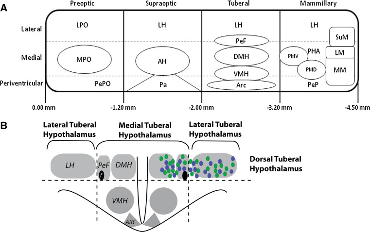
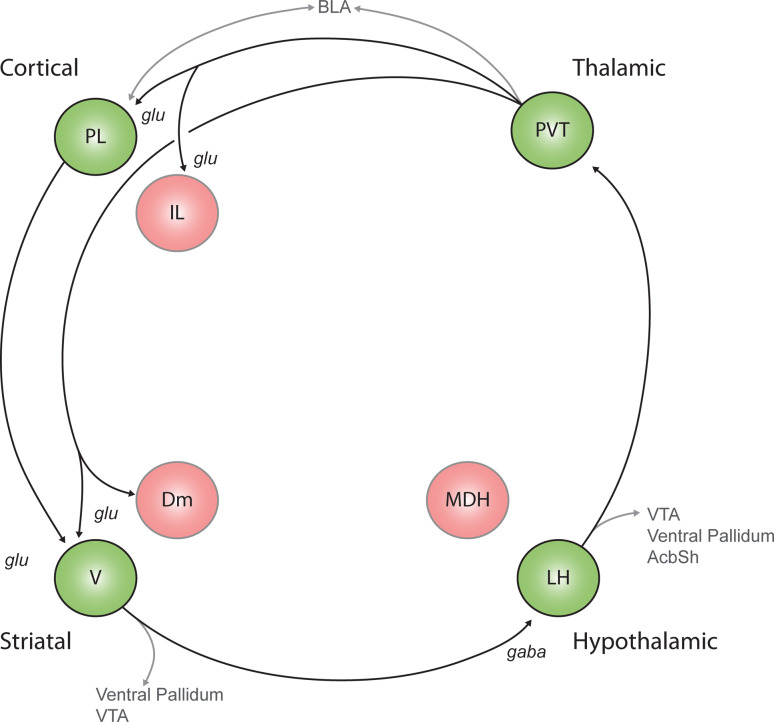
The hypothalamus is a neural structure critical for expression of motivated behaviours that ensure survival of the individual and the species. It is a heterogeneous structure, generally recognised to have four distinct regions in the rostrocaudal axis (preoptic, supraoptic, tuberal and mammillary). The tuberal hypothalamus in particular has been implicated in the neural control of appetitive motivation, including feeding and drug seeking. Here we review the role of the tuberal hypothalamus in appetitive motivation. First, we review evidence that different regions of the hypothalamus exert opposing control over feeding. We then review evidence that a similar bi-directional regulation characterises hypothalamic contributions to drug seeking and reward seeking. Lateral regions of the dorsal tuberal hypothalamus are important for promoting reinstatement of drug seeking, whereas medial regions of the dorsal tuberal hypothalamus are important for inhibiting this drug seeking after extinction training. Finally, we review evidence that these different roles for medial versus lateral dorsal tuberal hypothalamus in promoting or preventing reinstatement of drug seeking are mediated, at least in part, by different populations of hypothalamic neurons as well as the neural circuits in which they are located.


Figures

Fig. 1
Fig. 1
Highly simplified schematic outlining the anatomical organisation of the hypothalamus. a Schematic representation of the four rostrocaudal regions and three mediolateral zones of the hypothalamus. This figure is adapted from Fig. 1 of [2]. A large number of the subnuclei of the preoptic and supraoptic regions are omitted for clarity, as they are not discussed within the text of this review. Numbers represent mm from the bregma. b Schematic of a coronal section of the tuberal hypothalamus depicting the anatomical organisation of the tuberal hypothalamus discussed within this review. Blue circles represent the distribution of orexin neurons, and green circles represent the distribution of MCH neurons. ARC arcuate nucleus, AH anterior hypothalamic nucleus, DMH dorsomedial nucleus, f fornix, LH lateral hypothalamus, LM, lateral mammillary nucleus, LPO lateral preoptic area, MM medial mammillary nucleus, MPO medial preoptic nucleus, Pa paraventricular nucleus, PeF perifornical nucleus, PeP, posterior periventricular, PePO preoptic periventricular nucleus, PHA posterior hypothalamic area, PMD dorsal premammillary nucleus, PMV ventral premammillary nucleus, SuM, supramammillary nucleus, VMH ventromedial nucleus
Fig. 2
Fig. 2
A model depicting the neural circuitry associated with reinstatement of extinguished drug and reward seeking. The promotion of reward seeking is proposed to be mediated in large part by a circuit comprising the prelimbic prefrontal cortext (PL), ventral nucleus accumbens shell (V), lateral hypothalamus (LH) and paraventricular thalamus (PVT). We are proposing that the continued activation of this cortical-striatal-hypothalamic-thalamic-cortical “loop” is important for drug seeking during reinstatement. Multiple outputs from this loop, such as to the ventral pallidum and brainstem, enable expression of behaviour. Functional inhibition of all of these structures attenuates reinstatement of reward seeking, indicating that each node of this circuit is required to promote reward seeking. Evidence for direct V→LH and PVT→V pathways is reviewed in Sect. 3.1. Connections with this circuit and other structures implicated in these behaviours are displayed in grey. AcbSh nucleus accumbens shell, BLA basolateral amygdala, Dm dorsomedial accumbens shell, glu glutamate, IL infralimbic prefrontal cortex, MDH medial dorsal hypothalamus, VTA ventral tegmental area
Fig. 3
Fig. 3
A model depicting the neural circuitry associated with the expression of extinction of drug and reward seeking. The key feature of this model is that this circuit inhibits reward seeking via inhibition of activity in the cortical-striatal-hypothalamic-thalamic-cortical loop promoting reinstatement. The extinction circuit converges onto the reward-seeking circuit at two points, the lateral hypothalamus (LH) and paraventricular thalamus (PVT). Evidence for direct Dm→LH and MDH→PVT pathways is reviewed in Sect. 3.2. The AcbSh nucleus accumbens shell, BLA basolateral amygdala, Dm dorsomedial accumbens shell, glu glutamate, IL infralimbic prefrontal cortex, LHa lateral hypothalamus anterior, MDH medial dorsal hypothalamus, VTA ventral tegmental area

Similar articles

Cited by

References

    1. Swanson LW. Cerebral hemisphere regulation of motivated behavior. Brain Res. 2000;886(1–2):113–164. doi: 10.1016/S0006-8993(00)02905-X. - DOI - PubMed
    1. Simerly RB. Anatomical substrates of hypothalamic integration. In: George P, editor. The rat nervous system. 3. Burlington: Academic Press; 2004. pp. 335–368.
    1. Olds J, Milner P. Positive reinforcement produced by electrical stimulation of septal area and other regions of rat brain. J Comp Physiol Psychol. 1954;47(6):419–427. doi: 10.1037/h0058775. - DOI - PubMed
    1. Stellar E. The physiology of motivation. Psychol Rev. 1954;61(1):5–22. doi: 10.1037/h0060347. - DOI - PubMed
    1. Elmquist JK, Elias CF, Saper CB. From lesions to leptin: hypothalamic control of food intake and body weight. Neuron. 1999;22(2):221–232. doi: 10.1016/S0896-6273(00)81084-3. - DOI - PubMed

Publication types

MeSH terms

LinkOut - more resources