. 2012 Mar;26(3):616-26.
doi: 10.1007/s00464-011-1920-x. Epub 2011 Sep 23.

Value of multidetector computed tomography image segmentation for preoperative planning in general surgery

Affiliations

Value of multidetector computed tomography image segmentation for preoperative planning in general surgery

Vincenzo Ferrari et al. Surg Endosc. 2012 Mar.

Abstract

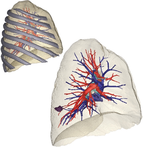
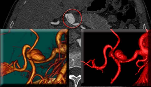
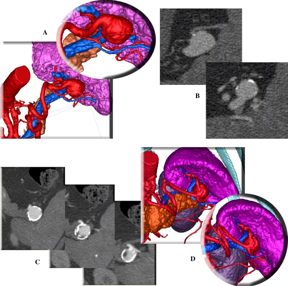
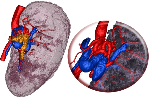
Background: Using practical examples, this report aims to highlight the clinical value of patient-specific three-dimensional (3D) models, obtained segmenting multidetector computed tomography (MDCT) images, for preoperative planning in general surgery.

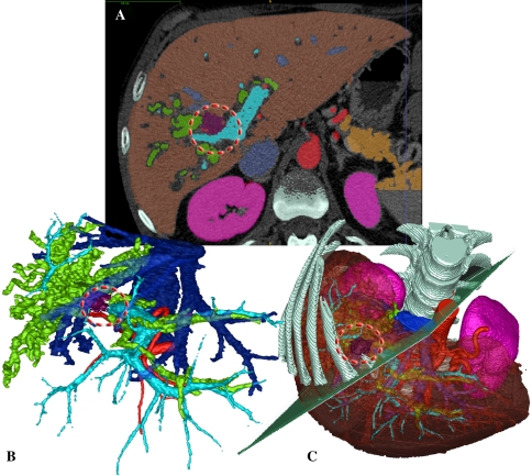
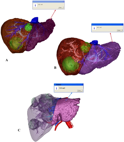
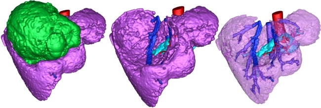
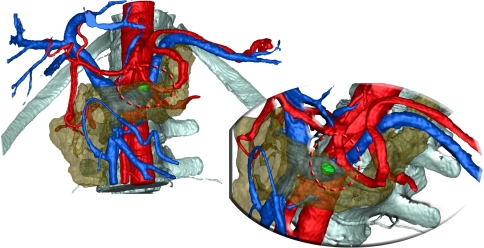
Methods: In this study, segmentation and 3D model generation were performed using a semiautomatic tool developed in the authors' laboratory. Their segmentation procedure is based on the neighborhood connected region-growing algorithm that, appropriately parameterized for the anatomy of interest and combined with the optimal segmentation sequence, generates good-quality 3D images coupled with facility of use. Using a touch screen monitor, manual refining can be added to segment structures unsuitable for automatic reconstruction. Three-dimensional models of 10 candidates for major general surgery procedures were presented to the operating surgeons for evaluation. A questionnaire then was administered after surgery to assess the perceived added value of the new technology.

Results: The questionnaire results were very positive. The authors recorded the diffuse opinion that planning the procedure using a segmented data set allows the surgeon to plan critical interventions with better awareness of the specific patient anatomy and consequently facilitates choosing the best surgical approach.

Conclusions: The benefit shown in this report supports a wider use of segmentation software in clinical practice, even taking into account the extra time and effort required to learn and use these systems.


Figures

Fig. 1
Fig. 1
A Computed tomography (CT) slice with superimposed segmentation. Note the tumor positioning. B, C Three-dimensional (3D) rendering of the segmented data set. Structures can be removed or made transparent for exact visualization of tumor positioning and tumor relationship with vessels and biliar ducts
Fig. 2
Fig. 2
A First segmented model showing liver vessels, metastases, and parenchyma. The residual healthy volume is estimated. B Three-dimensional (3D) model 6 weeks after embolization of the right portal system. Vasculature metastasis, embolized vessels, and parenchyma are rendered. The residual healthy volume is 45% larger. C A similar case after embolization in which the embolization did not achieve the hoped hypertrophy of the left lobe
Fig. 3
Fig. 3
Large epatic lesion that involves most of the upper liver segments. Beyond the tumor, it can be seen that the vascular system is only marginally involved and that the tumor can be safely enucleated
Fig. 4
Fig. 4
Three-dimensional (3D) model showing the little lesion in the pancreas parenchyma, the path of the vessels, and their positions
Fig. 5
Fig. 5
Three-dimensional (3D) model of the lung with and without the rib cage. The position of the ribs is important for planning the insertion points for the trocars. On the other hand, the transparent image facilitates understanding concerning the position of the tumor and its relationship with the hilar structures
Fig. 6
Fig. 6
Left direct volume rendering of an aneurysm close to the bifurcation of the common hepatic artery involving the proper hepatic artery and the gastroduodenal artery. This image does not permit an understanding of its origin. Right three-dimensional (3D) model obtained through segmentation, which shows exactly that the aneurysm involves only the common hepatic artery. In the background, one of the computed tomography (CT) slices can be seen with the aneurysm highlighted in the red circle
Fig. 7
Fig. 7
Two cases of splenic artery aneurysm. (A, B), the three- dimensional (3D) model, and the corresponding computed tomogra- phy (CT) slices for case 8. C The CT slices for case 9 show the ambiguity of afferent and efferent vessels. D The corresponding 3D model with and without the pancreatic tail facilitates understanding of the vascular anatomy and its anatomic relationship
Fig. 8
Fig. 8
Three-dimensional (3D) model of a megaspleen. It is possible precisely to understand the topology of vessels and to delineate the deformed pancreatic tail

References

    1. Dawes TJ, Vowler SL, Allen CM, Dixon AK. Training improves medical student performance in image interpretation. Br J Radiol. 2004;77:775–776. doi: 10.1259/bjr/66388556. - DOI - PubMed
    1. Fishman EK, Ney DR, Heath DG, Corl FM, Horton KM, Johnson PT. Volume rendering versus maximum intensity projection in CT angiography: what works best, when, and why. Radiographics. 2006;26:905–922. doi: 10.1148/rg.263055186. - DOI - PubMed
    1. Ishikawa T, UshikI T, Mizuno K, Togashi T, Watanabe K, Seki K, Ohta H, Yoshida T, Takeda K, Kamimura T. CT-maximum intensity projection is a clinically useful modality for the detection of gastric varices. World J Gastroenterol. 2005;11:7515–7519. - PMC - PubMed
    1. Mori G, Tokunaga D, Takahashi KA, Hojo T, Fujiwara H, Arai Y, Taniguchi D, Takatori R, Imai K, Otakara E, Ito H, Nishimura T, Kubo T. Maximum intensity projection as a tool to diagnose early rheumatoid arthritis. Mod Rheumatol. 2008;18:247–251. doi: 10.1007/s10165-008-0043-2. - DOI - PubMed
    1. Valencia R, Denecke T, Lehmkuhl L, Fischbach F, Felix R, Knollmann F. Value of axial and coronal maximum intensity projection (MIP) images in the detection of pulmonary nodules by multislice spiral CT: comparison with axial 1-mm and 5-mm slices. Eur Radiol. 2006;16:325–332. doi: 10.1007/s00330-005-2871-1. - DOI - PubMed

Publication types