

Reconstruction plate versus minimal invasive retrograde titanium elastic nail fixation for displaced midclavicular fractures
- PMID: 21948051
- PMCID: PMC3225608
- DOI: 10.1007/s10195-011-0158-7
Reconstruction plate versus minimal invasive retrograde titanium elastic nail fixation for displaced midclavicular fractures
Abstract
Background: Nonoperative management of midshaft clavicle fractures (MSCFs) is standard; however, it is now generally accepted that displaced midshaft clavicle fractures benefit from internal fixation. Plating and intramedullary fixation have become the accepted methods of fixation. The purpose of this study was to see if one method of fixation of clavicle fractures has a lower complication rate and higher union rate than the other.
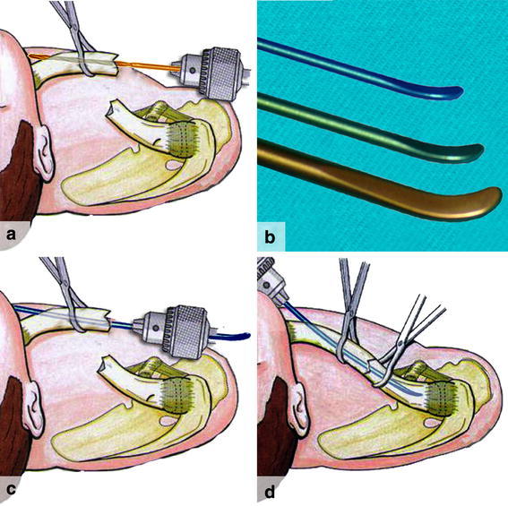
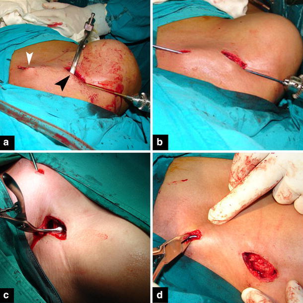
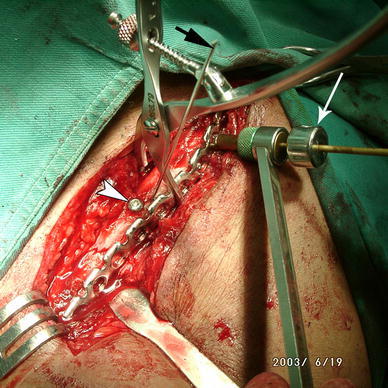
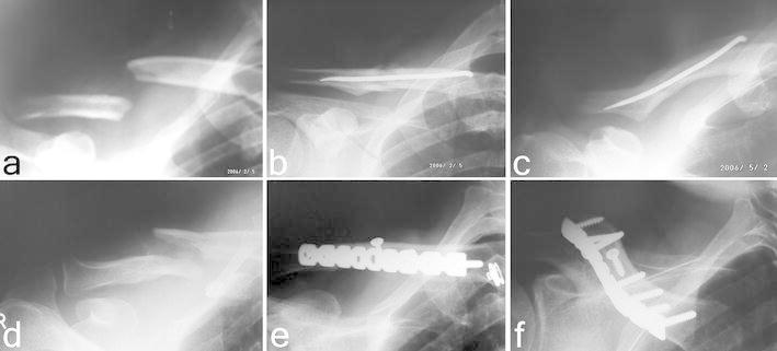
Materials and methods: Between December 2003 and September 2008, 38 patients were treated randomly by either plating (plate group) or retrograde nailing (RTEN group). Primary outcome measures included functional Constant scores, radiological union rate and union time. Clinical and radiological assessments were performed at the 6th week and the 3rd, 6th and 12th month postoperatively. Secondary outcome measures included the perioperative data (mean surgery time, blood loss, wound size, and hospital stay), and the complication rates.
Results: Similar results were found between the two groups regarding functional and radiological outcome after the 12th week (P > 0.05). However, earlier union and functional recovery were obtained at the 6th week for the RTEN group (P < 0.05). The rate of complications was significantly higher (15.8%) in the plate group compared with the RTEN group (0%; P > 0.05). In the plate group, significantly higher values were obtained for the perioperative data (P < 0.001).
Conclusion: Both techniques are equally effective at treating displaced midclavicular fractures, and give better function and fewer complications than nonoperative treatment. The RTEN technique has more advantages and lower complications than plating, making its use more favorable. It is recommended for athletes and young active individuals, and can be used as an alternative to conservative treatment or plate fixation.
Figures
References
-
- Ali KM, Lucas HK. Plating of fractures of the middle third of the clavicle. Injury. 1978;9(4):263–267. - PubMed
-
- Canadian Orthopaedic Trauma Society (2007) Nonoperative treatment compared with plate fixation of displaced midshaft clavicular fractures. A multicenter, randomized clinical trial. J Bone Joint Surg 89-A:1–10 - PubMed
-
- Collinge C, Devinney S, Herscovici D, DiPasquale T, Sanders R (2006) Anterior-inferior plate fixation of middle-third fractures and nonunions of the clavicle. J Orthop Trauma 20(10):680–686 - PubMed
-
- Constant CR, Murley AG. A clinical method of functional assessment of the shoulder. Clin Orthop Relat Res. 1987;214:160–164. - PubMed
Publication types
MeSH terms
Substances
LinkOut - more resources
Full Text Sources
Medical
