. 2012 May;470(5):1498-506.
doi: 10.1007/s11999-011-2070-0. Epub 2011 Sep 24.

Epithelioid hemangioma of bone and soft tissue: a reappraisal of a controversial entity

Affiliations

Epithelioid hemangioma of bone and soft tissue: a reappraisal of a controversial entity

Costantino Errani et al. Clin Orthop Relat Res. 2012 May.

Abstract

Background: The controversy surrounding diagnosis of an epithelioid hemangioma (EH), particularly when arising in skeletal locations, stems not only from its overlapping features with other malignant vascular neoplasms, but also from its somewhat aggressive clinical characteristics, including multifocal presentation and occasional lymph node involvement. Specifically, the distinction from epithelioid hemangioendothelioma (EHE) has been controversial. The recurrent t(1;3)(p36;q25) chromosomal translocation, resulting in WWTR1-CAMTA1 fusion, recently identified in EHE of various anatomic sites, but not in EH or other epithelioid vascular neoplasms, suggests distinct pathogeneses.

Question/purposes: We investigated the clinicopathologic and radiologic characteristics of bone and soft tissue EHs in patients treated at our institution with available tissue for molecular testing.

Patients and methods: Seventeen patients were selected after confirming the pathologic diagnosis and fluorescence in situ hybridization analysis for the WWTR1 and/or CAMTA1 rearrangements. Four patients had multifocal presentation. Most patients with EH of bone were treated by intralesional curettage. None of the patients died of disease and only four patients had a local recurrence.

Results: Our results, using molecular testing to support the pathologic diagnosis of EH, reinforce prior data that EH is a benign lesion characterized by an indolent clinical course with an occasional multifocal presentation and rare metastatic potential to locoregional lymph nodes.

Conclusion: These findings highlight the importance of distinguishing EH from other malignant epithelioid vascular tumors as a result of differences in their management and clinical outcome.

Level of evidence: Level IV, prognostic study. See Guidelines for Authors for a complete description of levels of evidence.


Figures

Fig. 1
Fig. 1
Three-color fluorescence in situ hybridization (FISH) shows no break-apart (split signal) in the region of 3q25.1-25.3; orange (RP11-690M20, RP11-718M13); green (RP11-286N6, RP11-157D5, RP11-64O23); or red (RP11-615G7, RP11-152K5, RP11-880C10).
Fig. 2A–B
Fig. 2A–B
(A) A radiograph shows the first metatarsal has been replaced and expanded by a multiseptated lytic lesion. No gross calcified matrix is evident in the lesion. An interrupted periosteal reaction was present on the radiograph obtained 16 months earlier (not shown) when the lesion was much smaller. (B) A coronal noncontrast CT image through the forefoot of the same patient shows marked expansion of the first metatarsal with extensive cortical destruction and several thin intralesional septa. The attenuation of the tumor is slightly lower than that of muscle.
Fig. 3A–B
Fig. 3A–B
(A) The en bloc resection specimen shows a diffusely hemorrhagic cut surface lesion expanding the rib. (B) A contrast-enhanced CT image of the same patient shows a multiseptated, expansile lytic lesion in the anterior portion of the right ninth rib, indenting and causing low-attenuation presumed to be reactive edema in the subjacent liver.
Fig. 4
Fig. 4
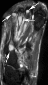
An axial T2-weighted MR image shows tumor deposits as areas of bright signal in several bones of foot (arrows).
Fig. 5A–C
Fig. 5A–C
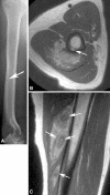
(A) The radiograph shows a tiny permeative, lytic focus in the posterior cortex of the midhumeral shaft (arrow). No calcifications are evident in nearby soft tissues. (B) An axial proton density MR image shows heterogeneous tumor deposits in the triceps muscle and an intracortical tumor deposit (arrow). (C) A sagittal proton density MR image shows multinodular tumor deposits with low-signal intensity inner rings (arrows), possibly related to hemosiderin deposition.
Fig. 6A–I
Fig. 6A–I
Histologic analysis showed (A) a lobular growth pattern of the lesion with extension outside the cortex in adjacent soft tissue (Stain, hematoxylin & eosin; original magnification, ×40); (B) mature vascular lumen formation with eosinophils infiltrating the adjacent stroma (Stain, hematoxylin & eosin; original magnification, ×100); (C) vasoformative properties at the periphery of the lesion, with larger caliber vessels lined by epithelioid cells (Stain, hematoxylin & eosin; original magnification, ×100); (D) hobnailed endothelial cells protruding in the lumen in a characteristic ‘tombstone’ appearance (Stain, hematoxylin & eosin; original magnification, ×100); (E) the central portion of the lesion typically had more solid growth with sheets of epithelioid cells with densely eosinophilic cytoplasm and lacked obvious vessel formation. Occasionally abundant erythrocyte extravasation was seen (Stain, hematoxylin & eosin; original magnification, ×200); (F) epithelioid cells with a more foamy, vacuolated cytoplasm and focal, moderate pleomorphism and pseudonuclear inclusions (Stain, hematoxylin & eosin; original magnification, ×400); (G) intracytoplasmic vacuoles but typically these were not a predominant feature (Stain, hematoxylin & eosin; original magnification, ×200); (H) occasional areas of a bland spindle cell component (Stain, hematoxylin & eosin; original magnification, ×200); and (I) vascular invasion in one patient who had lymph node spread (Stain, hematoxylin & eosin; original magnification, ×100).

References

    1. Antonescu CR, Zhang L, Chang NE, Pawel BR, Travis W, Katabi N, Edelman M, Rosenberg AE, Nielsen GP, Dal Cin P, Fletcher CD. EWSR1-POU5F1 fusion in soft tissue myoepithelial tumors: a molecular analysis of sixty-six cases, including soft tissue, bone, and visceral lesions, showing common involvement of the EWSR1 gene. Genes Chromosomes Cancer. 2010;49:1114–1124. doi: 10.1002/gcc.20819. - DOI - PMC - PubMed
    1. Deshpande V, Rosenberg AE, O’Connell JX, Nielsen GP. Epithelioid angiosarcoma of the bone: a series of 10 cases. Am J Surg Pathol. 2003;27:709–716. doi: 10.1097/00000478-200306000-00001. - DOI - PubMed
    1. Deyrup AT, Montag AG. Epithelioid and epithelial neoplasms of bone. Arch Pathol Lab Med. 2007;131:205–216. - PubMed
    1. Deyrup AT, Tighiouart M, Montag AG, Weiss SW. Epithelioid hemangioendothelioma of soft tissue: a proposal for risk stratification based on 49 cases. Am J Surg Pathol. 2008;32:924–927. doi: 10.1097/PAS.0b013e31815bf8e6. - DOI - PubMed
    1. Errani C, Zhang L, Sung SY, Hajdu M, Singer S, Maki RG, Healey JH, Antonescu CR. A novel WWTR1-CAMTA1 gene fusion is a consistent abnormality in epithelioid hemangioendothelioma of different anatomic sites. Genes Chromosomes Cancer. 2011;50:644–653. doi: 10.1002/gcc.20886. - DOI - PMC - PubMed

Publication types

MeSH terms

Substances