. 2011 Oct 17;52(11):8216-23.
doi: 10.1167/iovs.11-8213.

Human corneal endothelial cells employ phosphorylation of p27(Kip1) at both Ser10 and Thr187 sites for FGF-2-mediated cell proliferation via PI 3-kinase

Affiliations

Human corneal endothelial cells employ phosphorylation of p27(Kip1) at both Ser10 and Thr187 sites for FGF-2-mediated cell proliferation via PI 3-kinase

Jeong Goo Lee et al. Invest Ophthalmol Vis Sci. .

Abstract

Purpose: FGF-2 stimulates cell proliferation of rabbit corneal endothelial cells (rCECs) by degrading the cyclin-dependent kinase inhibitor p27(Kip1) (p27) through its phosphorylation mechanism. The authors investigated whether the cell proliferation of human CECs (hCECs) is also induced by FGF-2 stimulation through the p27 phosphorylation pathway.

Methods: Expression and activation of protein were analyzed by immunoblotting. Cell proliferation was measured by 3-(4,5-dimethylthiazolyl-2)-2,5-diphenyltetrazolium bromide (MTT) assay. Transfection of hCECs with small interference RNA (siRNA) was performed using a transfection reagent.

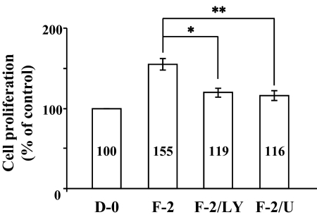
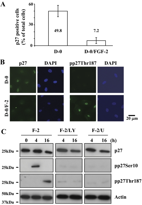
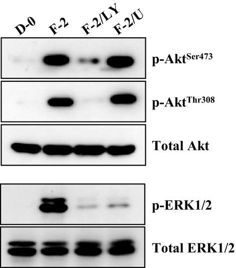
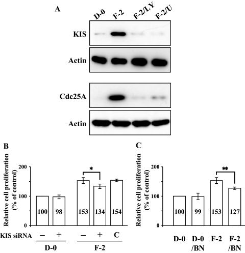
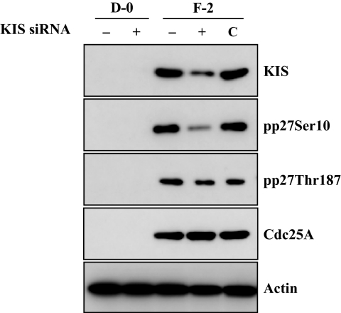
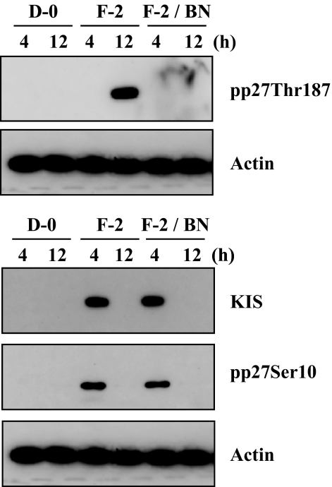
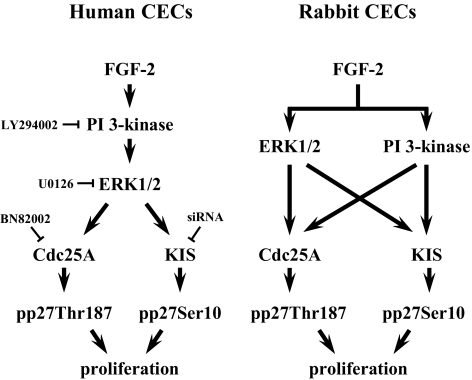
Results: FGF-2 stimulated cell proliferation in hCECs; the FGF-2 action was completely blocked by pathway-specific inhibitors for PI 3-kinase (LY294002) and MEK1/2 (U0126), respectively. Using immunoblotting, the authors showed that FGF-2 induced phosphorylation of p27 at both serine 10 (Ser10) and threonine 187 (Thr187) sites. These effects were also completely blocked by LY294002 or U0126. The authors then determined cross-talk between PI 3-kinase and extracellular signal-regulated kinase (ERK)1/2; blocking of ERK1/2 activation by LY294002 indicated that in hCECs ERK1/2 works as a downstream effector to PI 3-kinase for cell proliferation induced by FGF-2, whereas the ERK1/2 pathway in rCECs is parallel to the PI 3-kinase pathway. However, the downstream mechanism involved in cell cycle progression in hCECs is identical to that of rCECs: phosphorylation of p27 at Ser10 was mediated by kinase-interacting stathmin (KIS), confirmed with siRNA to KIS, and phosphorylation of p27 at Thr187 was mediated by cell division cycle 25A (Cdc25A), confirmed using Cdc25A inhibitor. CONCLUSIONS; FGF-2 stimulates proliferation of hCECs through PI 3-kinase and its downstream target ERK1/2 pathways. This linear signal transduction significantly downregulates p27 through its phosphorylation at both Ser10 and Thr187 sites mediated by KIS and Cdc25A, respectively.


Figures

Figure 1.
Figure 1.
Inhibitory effect of PI 3-kinase and ERK1/2 inhibitor on cell proliferation stimulated by FGF-2. Cell proliferation was determined by MTT assay. The serum-starved hCECs were pretreated with LY294002 for PI 3-kinase inhibition or U0126 for ERK1/2 inhibition for 2 hours and then maintained in DMEM with FGF-2 for 24 hours. At the end of incubation, MTT was added for 4 hours and then intracellular purple formazan, the MTT metabolic product by the action of dehydrogenase enzymes of metabolically-active cells, was quantified with a spectrophotometric plate reader at dual wavelengths of 570 and 650 nm. Data were normalized to cells maintained in DMEM without serum. D-0, DMEM without serum; F-2, FGF-2; LY, LY294002; U, U0126. The graphs represent the mean ± SEM from three independent experiments. (*P = 0.0009, paired t-test; **P = 0.0002, paired t-test).
Figure 2.
Figure 2.
Reduction of p27 level ex vivo and induction of its phosphorylation at both Ser10 and Thr187 sites by FGF-2 stimulation in vitro. (A) The corneal endothelium was incubated with or without FGF-2 for 24 hours and then immunostained with anti-p27 antibody. The total DAPI-stained and p27-positive cells were counted in one microscopic field under high magnification (×400). The percentage of p27-positive CECs was significantly greater in the corneal pieces incubated in mitogen-deprived medium (D-0) than in those treated with FGF-2. (B) The serum-starved hCECs were treated with or without FGF-2 for 16 hours and maintained in D-0 for up to 24 hours. The cultured cells were fixed and labeled with anti-p27 or anti-pp27Thr187 antibody (FITC) and DAPI, respectively. (C) The serum-starved hCECs were pretreated with PI 3-kinase inhibitor or MEK1/2 inhibitor for 2 hours and then maintained in DMEM with FGF-2 for the designated time. At the end of treatment, cells were lysed and then immunoblotted with the respective antibody. Actin was used to control protein concentration on immunoblot analysis. The results represent data obtained in three independent experiments.
Figure 3.
Figure 3.
Activation of ERK1/2 by FGF-2 stimulation through PI 3-kinase. The serum-starved hCECs were pretreated with PI 3-kinase inhibitor or MEK1/2 inhibitor for 2 hours and then maintained in DMEM with FGF-2 for 8 hours. At the end of treatment, cells were lysed and then immunoblotted with the designated antibody. ERK1/2 works as a downstream regulator to the PI 3-kinase/Akt pathways triggered by FGF-2 stimulation. Total Akt and ERK1/2 were used to control protein concentration on immunoblot analysis. The results represent data obtained in three independent experiments.
Figure 4.
Figure 4.
Induction of KIS and Cdc25A through PI 3-kinase and ERK1/2 and their involvement in cell proliferation induced by FGF-2 stimulation. (A) The serum-starved hCECs were pretreated with PI 3-kinase inhibitor or MEK1/2 inhibitor for 2 hours and then maintained in DMEM with FGF-2 for 4 hours to detect KIS and 12 hours to detect Cdc25A. At the end of treatment, cells were lysed and then immunoblotted with the designated antibody. Actin was used to control protein concentration on immunoblot analysis. The results represent data obtained in three independent experiments. (B) After transfection with KIS siRNA, the CECs were maintained with DMEM or DMEM with FGF-2 for 24 hours. Cell proliferation was determined using MTT assay as previously described in Figure 1. Data were normalized to untransfected cells. The graphs represent the mean ± SEM from three independent experiments (*P = 0.0114, paired t-test). (C) To determine the effect of Cdc25A inhibition on cell proliferation, the serum-starved cells were pretreated with BN82002 for Cdc25A inhibition for 2 hours and then maintained in DMEM with or without FGF-2 for 24 hours. Cell proliferation was determined and data were normalized to unstimulated cells. The graphs represent the mean ± SEM from three independent experiments (**P = 0.0087, paired t-test). BN, BN82002.
Figure 5.
Figure 5.
Inhibitory effect of siRNA to KIS on phosphorylation of p27 at the Ser10 site, but not at the Thr187 site. Human CECs were transfected with KIS siRNA. After transfection, cells were maintained with DMEM or DMEM with FGF-2 for an additional 24 hours. Total protein extracts were prepared and immunoblotted with designated antibodies. Actin was used to control protein concentration on immunoblot analysis. The results represent data obtained in three independent experiments. C, negative control cells which were transfected with negative control siRNA (Silencer Select Negative Control; Ambion).
Figure 6.
Figure 6.
Inhibitory effect of Cdc25A inhibitor on phosphorylation of p27 at the Thr187 site, but not at the Ser10 site. The serum-starved hCECs were pretreated with or without Cdc25A inhibitor for 2 hours and then maintained in DMEM with FGF-2 for the designated time. At the end of treatment, the phosphorylation of p27 at the Ser10 site and the Thr187 site was detected with immunoblotting. Actin was used to control protein concentration on immunoblot analysis. The results represent data obtained in three independent experiments.
Figure 7.
Figure 7.
Schematic comparison of human and rabbit CECs cell proliferation pathway stimulated by FGF-2. ERK1/2 pathway activated by PI 3-kinase increase cell proliferation through phosphorylation of p27 at both Ser10 and Thr187 residues in response to FGF-2 stimulation in hCECs. In contrast, parallel ERK1/2 and PI 3-kinase pathways induced by FGF-2 increase cell proliferation in rCECs.

References

    1. Kreutziger GO. Lateral membrane morphology and gap junction structure in rabbit corneal endothelium. Exp Eye Res. 1976;23:285–293 - PubMed
    1. Geroski DH, Edelhauser HF. Quantitation of Na/K ATPase pump sites in the rabbit corneal endothelium. Invest Ophthalmol Vis Sci. 1984;25:1056–1060 - PubMed
    1. Joyce NC. Proliferative capacity of the corneal endothelium. Prog Retin Eye Res. 2003;22:359–389 - PubMed
    1. Joyce NC, Navon SE, Roy S, Zieske JD. Expression of cell cycle-associated proteins in human and rabbit corneal endothelium in situ. Invest Ophthalmol Vis Sci. 1996;37:1566–1575 - PubMed
    1. Senoo T, Joyce NC. Cell cycle kinetics in corneal endothelium from old and young donors. Invest Ophthalmol Vis Sci. 2000;41:660–667 - PubMed

Publication types

MeSH terms