. 2011 Nov;77(22):8154-63.
doi: 10.1128/AEM.05735-11. Epub 2011 Sep 23.

4,6-α-glucanotransferase, a novel enzyme that structurally and functionally provides an evolutionary link between glycoside hydrolase enzyme families 13 and 70

Affiliations

4,6-α-glucanotransferase, a novel enzyme that structurally and functionally provides an evolutionary link between glycoside hydrolase enzyme families 13 and 70

Slavko Kralj et al. Appl Environ Microbiol. 2011 Nov.

Abstract

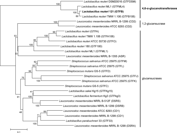
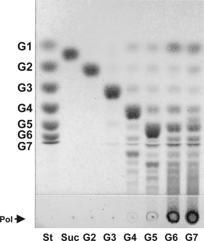
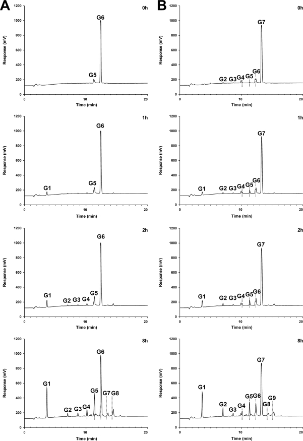
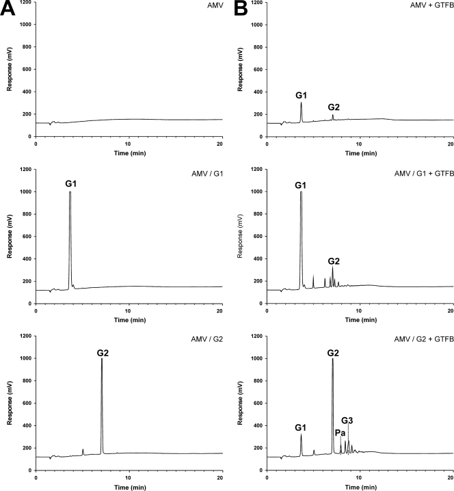
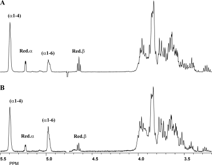
Lactobacillus reuteri 121 uses the glucosyltransferase A (GTFA) enzyme to convert sucrose into large amounts of the α-D-glucan reuteran, an exopolysaccharide. Upstream of gtfA lies another putative glucansucrase gene, designated gtfB. Previously, we have shown that the purified recombinant GTFB protein/enzyme is inactive with sucrose. Various homologs of gtfB are present in other Lactobacillus strains, including the L. reuteri type strain, DSM 20016, the genome sequence of which is available. Here we report that GTFB is a novel α-glucanotransferase enzyme with disproportionating (cleaving α1→4 and synthesizing α1→6 and α1→4 glycosidic linkages) and α1→6 polymerizing types of activity on maltotetraose and larger maltooligosaccharide substrates (in short, it is a 4,6-α-glucanotransferase). Characterization of the types of compounds synthesized from maltoheptaose by matrix-assisted laser desorption ionization-time of flight mass spectrometry (MALDI-TOF MS), methylation analysis, and 1-dimensional ¹H nuclear magnetic resonance (NMR) spectroscopy revealed that only linear products were made and that with increasing degrees of polymerization (DP), more α1→6 glycosidic linkages were introduced into the final products, ranging from 18% in the incubation mixture to 33% in an enriched fraction. In view of its primary structure, GTFB clearly is a member of the glycoside hydrolase 70 (GH70) family, comprising enzymes with a permuted (β/α)₈ barrel that use sucrose to synthesize α-D-glucan polymers. The GTFB enzyme reaction and product specificities, however, are novel for the GH70 family, resembling those of the GH13 α-amylase type of enzymes in using maltooligosaccharides as substrates but differing in introducing a series of α1→6 glycosidic linkages into linear oligosaccharide products. We conclude that GTFB represents a novel evolutionary intermediate between the GH13 and GH70 enzyme families, and we speculate about its origin.


Figures

Fig. 1.
Fig. 1.
Unrooted phylogenetic tree of GTF proteins (glucansucrases and [putative] 4,6-α-glucanotransferases) from lactic acid bacteria. Alignments and dendrogram construction were carried out (using the catalytic cores only [for example, from “WYRP” to “WVPDQ” of L. reuteri 121 GTFA]) with MEGA version 4, using the neighbor joining method. Bootstrap values (expressed as percentages) are given at the branching points. The bar corresponds to a genetic distance of 0.1 substitution per position (10% amino acid sequence difference).
Fig. 2.
Fig. 2.
Amino acid sequence (http://www.cazy.org) alignment of conserved regions (II, III, IV, and I) in the catalytic domains of (putative) 4,6-α-glucanotransferase enzymes (A), DSRE (5) and DSRP, glucansucrase enzymes containing two catalytic domains (CD1 and CD2) (B), and dextran-, mutan-, alternan-, and reuteransucrase enzymes of lactic acid bacteria (C) (see also references and 23). The seven strictly conserved amino acid residues (indicated by the numbers 1 to 7 above the sequences; underlined and lightly shaded in L. reuteri 121 GTFA and GTFB), with important contributions to the −1 and +1 subsites in glucansucrase enzymes, are also conserved in the 4,6-α-glucanotransferase enzymes. GTFB amino acid D1015 (the putative nucleophilic residue), targeted in this study, is shown in boldface. Dark shading indicates changes in conserved amino acid residues between 4,6-α-glucanotransferases and glucansucrases; the corresponding amino acid numbering is indicated. *, low activity.
Fig. 3.
Fig. 3.
TLC analysis of the reaction products of 90 nM GTFB incubated for 13 h in 50 mM sodium acetate buffer, pH 4.7, containing 1 mM CaCl2 with 25 mM sucrose or 25 mM maltooligosaccharides. St, standard; Suc, sucrose; G1, glucose; G2, maltose; G3, maltotriose; G4, maltotetraose; G5, maltopentaose; G6, maltohexaose; G7, maltoheptaose; Pol, polymer.
Fig. 4.
Fig. 4.
HPAEC analysis of the reaction products of 90 nM GTFB incubated for 0 to 8 h in 50 mM sodium acetate buffer, pH 4.7, containing 1 mM CaCl2 with 25 mM maltohexaose (A) or 25 mM maltoheptaose (B).
Fig. 5.
Fig. 5.
HPAEC analysis of samples with 0.25% amylose-V (AMV) alone (donor substrate) or amylose-V with 25 mM glucose or 25 mM maltose (acceptor substrates), either without the GTFB enzyme (A) or with GTFB (90 nM) (B) incubated overnight at 37°C in 25 mM sodium acetate buffer, pH 4.7, containing 1 mM CaCl2. Pa, panose.
Fig. 6.
Fig. 6.
Five hundred-megahertz 1-dimensional 1H NMR analysis of maltoheptaose (DP7) incubated with 90 nM GTFB for 7 days in 50 mM sodium acetate buffer, pH 4.7, containing 1 mM CaCl2. (A) Incubation sample; (B) ethanol-precipitated fraction (P2) from the incubation sample. Red., reducing glucose unit.

References

    1. Argüello-Morales M. A., et al. 2000. Sequence analysis of the gene encoding alternansucrase, a sucrose glucosyltransferase from Leuconostoc mesenteroides NRRL B-1355. FEMS Microbiol. Lett. 182:81–85 - PubMed
    1. Ausubel F. M., et al. 1987. Current protocols in molecular biology. John Wiley & Sons, Inc., New York, NY
    1. Barends T. R. M., et al. 2007. Three-way stabilization of the covalent intermediate in amylomaltase, an α-amylase-like transglycosylase. J. Biol. Chem. 282:17242–17249 - PubMed
    1. Devulapalle K. S., Goodman S. D., Gao Q., Hemsley A., Mooser G. 1997. Knowledge-based model of a glucosyltransferase from the oral bacterial group of mutans streptococci. Protein Sci. 6:2489–2493 - PMC - PubMed
    1. Fabre E., et al. 2005. Role of the two catalytic domains of DSR-E dextransucrase and their involvement in the formation of highly α-1,2 branched dextran. J. Bacteriol. 187:296–303 - PMC - PubMed

MeSH terms

LinkOut - more resources