

Dietary ligands of the aryl hydrocarbon receptor induce anti-inflammatory and immunoregulatory effects on murine dendritic cells
- PMID: 21948866
- PMCID: PMC3216417
- DOI: 10.1093/toxsci/kfr249
Dietary ligands of the aryl hydrocarbon receptor induce anti-inflammatory and immunoregulatory effects on murine dendritic cells
Abstract
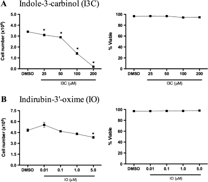
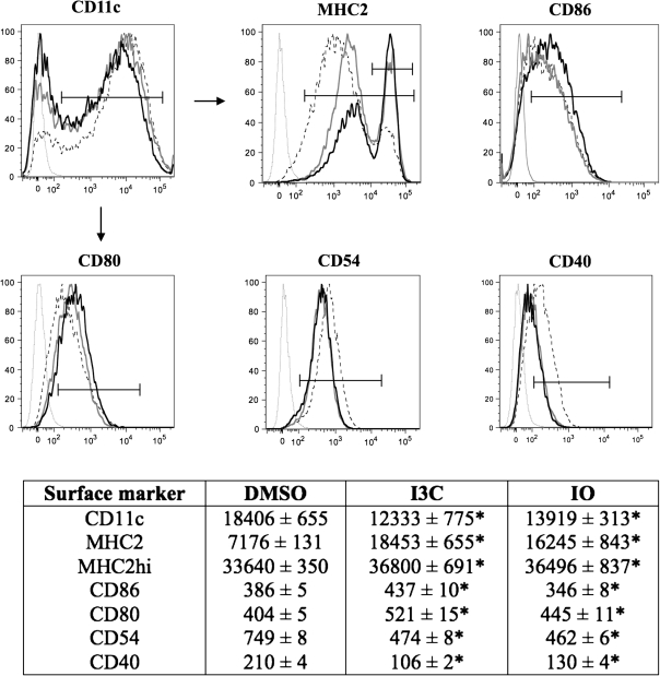
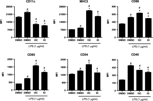
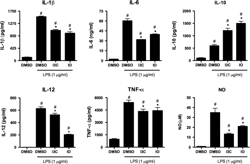
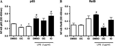
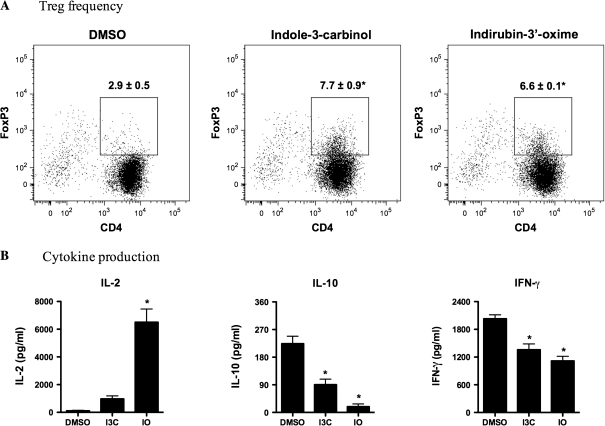
Activation of the aryl hydrocarbon receptor (AhR) in immune cells, such as dendritic cells (DCs), can lead to suppressed immune responses. Although AhR activation is most recognized for mediating the effects of its prototypical ligand 2,3,7,8-tetrachlorodibenzo-p-dioxin (TCDD), many compounds existing in dietary sources can also bind the AhR. Because the immunomodulatory effects of indole-3-carbinol (I3C) and indirubin-3'-oxime (IO) have yet to be investigated in DCs, we evaluated the potential immunomodulatory effects of these compounds on murine DCs. We hypothesized that I3C and IO suppress immune and inflammatory responses in DCs. We found that both I3C and IO decreased the expression of CD11c, CD40, and CD54 while they increased expression of MHC2 and CD80. Following lipopolysaccharide (LPS)-activation, I3C and IO suppressed the production of pro-inflammatory mediators including tumor necrosis factor-α, interleukin (IL)-1β, IL-6, IL-12, and nitric oxide but increased IL-10 levels. These effects of I3C and IO were partially mediated by the AhR. Additionally, immunoregulatory genes, such as ALDH1A, IDO and TGFB, were upregulated following treatment with I3C or IO. Both I3C and IO decreased basal levels of nuclear factor-kappa B p65, but only I3C suppressed the LPS-induced activity of RelB. Finally, when cultured with naïve T cells, bone marrow-derived dendritic cells treated with the dietary AhR ligands increased the frequency of Foxp3+ Tregs in an antigen-specific manner. Taken together, these results indicate that I3C and IO exhibit immunosuppressive and anti-inflammatory effects on DCs. Because I3C and IO are significantly less toxic than TCDD, these natural products may ultimately become useful therapeutics for the treatment of autoimmune and inflammatory diseases.
Figures
Similar articles
-
Consequences of AhR activation in steady-state dendritic cells.Toxicol Sci. 2011 Feb;119(2):293-307. doi: 10.1093/toxsci/kfq354. Epub 2010 Nov 19. Toxicol Sci. 2011. PMID: 21097750 Free PMC article.
-
Functional and phenotypic effects of AhR activation in inflammatory dendritic cells.Toxicol Appl Pharmacol. 2010 Jul;246(1-2):18-28. doi: 10.1016/j.taap.2010.03.013. Epub 2010 Mar 27. Toxicol Appl Pharmacol. 2010. PMID: 20350561 Free PMC article.
-
Immunoregulatory effects of indole-3-carbinol on monocyte-derived macrophages in systemic lupus erythematosus: A crucial role for aryl hydrocarbon receptor.Autoimmunity. 2018 Aug;51(5):199-209. doi: 10.1080/08916934.2018.1494161. Epub 2018 Oct 5. Autoimmunity. 2018. PMID: 30289282
-
Indole-3-carbinol induces tumor cell death: function follows form.J Surg Res. 2016 Jul;204(1):47-54. doi: 10.1016/j.jss.2016.04.021. Epub 2016 Apr 22. J Surg Res. 2016. PMID: 27451867 Free PMC article. Review.
-
The roles of aryl hydrocarbon receptor in immune responses.Int Immunol. 2013 Jun;25(6):335-43. doi: 10.1093/intimm/dxt011. Epub 2013 Apr 11. Int Immunol. 2013. PMID: 23580432 Review.
Cited by
-
Ambient urban dust particulate matter reduces pathologic T cells in the CNS and severity of EAE.Environ Res. 2019 Jan;168:178-192. doi: 10.1016/j.envres.2018.09.038. Epub 2018 Oct 1. Environ Res. 2019. PMID: 30316103 Free PMC article.
-
Environmental factors in autoimmune diseases and their role in multiple sclerosis.Cell Mol Life Sci. 2016 Dec;73(24):4611-4622. doi: 10.1007/s00018-016-2311-1. Epub 2016 Aug 4. Cell Mol Life Sci. 2016. PMID: 27491297 Free PMC article. Review.
-
Deletion of aryl hydrocarbon receptor AHR in mice leads to subretinal accumulation of microglia and RPE atrophy.Invest Ophthalmol Vis Sci. 2014 Aug 26;55(9):6031-40. doi: 10.1167/iovs.14-15091. Invest Ophthalmol Vis Sci. 2014. PMID: 25159211 Free PMC article.
-
Indole Propionic Acid, an Unusual Antibiotic Produced by the Gut Microbiota, With Anti-inflammatory and Antioxidant Properties.Front Microbiol. 2020 Oct 27;11:575586. doi: 10.3389/fmicb.2020.575586. eCollection 2020. Front Microbiol. 2020. PMID: 33193190 Free PMC article. Review.
-
Prenatal indole-3-carbinol administration activates aryl hydrocarbon receptor-responsive genes and attenuates lung injury in a bronchopulmonary dysplasia model.Exp Biol Med (Maywood). 2021 Mar;246(6):695-706. doi: 10.1177/1535370220963789. Epub 2020 Nov 4. Exp Biol Med (Maywood). 2021. PMID: 33148012 Free PMC article.
References
-
- Bradlow HL, Zeligs MA. Diindolylmethane (DIM) spontaneously forms from indole-3-carbinol (I3C) during cell culture experiments. In Vivo. 2010;24:387–391. - PubMed
-
- Chen YH, Dai HJ, Chang HP. Suppression of inducible nitric oxide production by indole and isothiocyanate derivatives from Brassica plants in stimulated macrophages. Planta Med. 2003;69:696–700. - PubMed
Publication types
MeSH terms
Substances
Grants and funding
LinkOut - more resources
Full Text Sources
Other Literature Sources
Research Materials
