. 2011 Nov 1;187(9):4907-12.
doi: 10.4049/jimmunol.1101519. Epub 2011 Sep 23.

Invariant NKT cell defects in vitamin D receptor knockout mice prevents experimental lung inflammation

Affiliations

Invariant NKT cell defects in vitamin D receptor knockout mice prevents experimental lung inflammation

Sanhong Yu et al. J Immunol. .

Abstract

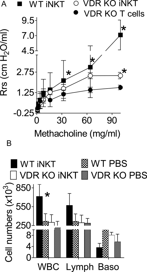
Vitamin D receptor (VDR) deficiency (knockout [KO]) results in a failure of mice to generate an airway hyperreactivity (AHR) response on both the BALB/c and C57BL/6 background. The cause of the failed AHR response is the defective population of invariant NKT (iNKT) cells in the VDR KO mice because wild-type (WT) iNKT cells rescued the AHR response. VDR KO mice had significantly fewer iNKT cells and normal numbers of T cells in the spleen compared with WT mice. In BALB/c VDR KO mice, the reduced frequencies of iNKT cells were not apparent in the liver or thymus. VDR KO and WT Th2 cells produced similar levels of IFN-γ and IL-5. On the BALB/c background, Th2 cells from VDR KO mice produced less IL-13, whereas on the C57BL/6 background, Th2 cells from VDR KO mice produced less IL-4. Conversely, VDR KO iNKT cells were defective for the production of multiple cytokines (BALB/c: IL-4, IL-5, and IL-13; C57BL/6: IL-4 and IL-17). Despite relatively normal Th2 responses, BALB/c and C57BL/6 VDR KO mice failed to develop AHR responses. The defect in iNKT cells as a result of the VDR KO was more important than the highly susceptible Th2 background of the BALB/c mice. Defective iNKT cell responses in the absence of the VDR result in the failure to generate AHR responses in the lung. The implication of these mechanistic findings for human asthma requires further investigation.


Figures

Figure 1
Figure 1. Balb/c VDR KO mice fail to develop experimental allergic asthma
Balb/c WT and VDRKO mice were induced to develop allergic asthma. A) AHR was measured in naïve WT (WT + PBS) and OVA/Alum immunized and OVA challenged WT and VDR KO mice. Immunized WT mice developed AHR following increasing doses of methacholine. Conversely, immunized VDR KO mice failed to respond and the AHR response was similar to the WT + PBS naïve mice. n= 4 mice per group and one representative experiment of 4. The WT AHR response was significantly higher than VDRKO or WT + PBS, *P<0.05. B) The histopathology section from one representative lung from each group of mice in A is shown. C) The mean histopathology scores from all of the sensitized mice in the group. Values from VDR KO mice are significantly different than WT mice (n=12 mice per group, mean ± SEM, *P<0.05).
Figure 2
Figure 2. αGalCer induced AHR fails in VDR KO mice on the Balb/c and C57BL/6 background
WT and VDRKO mice were challenged i.n. with αGalCer and analyzed 24h later. A) αGalCer induced sensitivity to methacoline in Balb/c and C57BL/6 WT mice. αGalCer treatment of either Balb/c or C57BL/6 VDR KO mice failed to induce an increase in air resistance. WT αGalCer AHR values are significantly higher than all other groups, *P<0.05 (1 representative of 2 experiments, n=3 mice per group per experiment). B) The histopathology section from one representative lung from each group of mice in A is shown. C) BALF from WT and VDRKO mice on the C57BL/6 mice were collected to determine the IL-4 level. IL-4 was detectable only in the WT BALF from mice exposed to αGalCer. WT αGalCer IL-4 values were significantly higher than all other groups, *P<0.05 (n=6 mice per group, error bars are ± SEM).
Figure 3
Figure 3. iNKT, CD4+ T, and CD8+ T cell numbers in the Balb/c VDR KO and WT mice
A) The total number of iNKT, CD4+ T and CD8+ T cells isolated from the spleen of Balb/c mice. B) The frequency of iNKT cells (CD1d tetramer/TCRβ+ T cells) in Balb/c WT and VDR KO spleen, thymus and liver. VDR KO mice had significantly fewer iNKT cells than WT in the spleen (n=4–6 mice per group). The frequency of iNKT, CD4+ T and CD8+ T cells isolated from the BALF of C) C57BL/6 and D) Balb/c VDR KO and WT mice (n=6–7 mice per group). E) The total number of iNKT cells in the BALF of the C57BL/6 and Balb/c mice.
Figure 4
Figure 4. Th2 and iNKT generated cytokine secretion from VDR KO and WT mice
Splenocytes from WT and VDRKO mice were stimulated under Th2 or iNKT cell conditions and the ability to produce IL-4, IL-5, IL-13, IL-17 and IFN-γ were evaluated. Th2 cell cultures of A) Balb/c or B) C57BL/6 spleens. iNKT cells cultures generated by αGalCer treatment of C) Balb/c or D) C57BL/6 splenocytes. Data shown are the mean ± SEM of 3–4 individual mice and 1 representative of 3 experiments.
Figure 5
Figure 5. WT iNKT cells rescued AHR in VDR KO mice
Mice were injected with αGalCer and immunized/challenged with OVA. VDR KO mice were given WT iNKT cells (VDR KO iNKT) or WT T cells depleted of iNKT cells (VDR KO T cells). A) WT mice developed AHR following exposure to methacholine and the AHR response was significantly higher than VDR KO that received T cells mice (at doses of methacholine over 16 mg/ml, n= 5 mice per group). VDR KO mice that received iNKT cells had higher AHR responses than VDR KO mice that received T cells and the difference was significant at 95 mg/ml methacholine (n=5 mice per group), * P<0.05. B) Cellular influx in the BALF of WT and VDRKO mice 24 h after last OVA i.n. challenge. WT iNKT - αGalCer and OVA sensitized, WT PBS- PBS and OVA sensitized, VDR KO iNKT, αGalCer /iNKT cell transfer and OVA sensitized, VDR KO PBS- PBS and OVA sensitized. The value from WT αGalCer mice were significantly higher than all other groups (n= 5 mice per group, *P<0.05).

References

    1. Wills-Karp M. Immunologic basis of antigen-induced airway hyperresponsiveness. Annu Rev Immunol. 1999;17:255–281. - PubMed
    1. Robinson DS, Hamid Q, Ying S, Tsicopoulos A, Barkans J, Bentley AM, Corrigan C, Durham SR, Kay AB. Predominant TH2-like bronchoalveolar T-lymphocyte population in atopic asthma. N Engl J Med. 1992;326:298–304. - PubMed
    1. Bendelac A, Savage PB, Teyton L. The biology of NKT cells. Annu Rev Immunol. 2007;25:297–336. - PubMed
    1. Akbari O, Stock P, Meyer E, Kronenberg M, Sidobre S, Nakayama T, Taniguchi M, Grusby MJ, DeKruyff RH, Umetsu DT. Essential role of NKT cells producing IL-4 and IL-13 in the development of allergen-induced airway hyperreactivity. Nat Med. 2003;9:582–588. - PubMed
    1. Pichavant M, Goya S, Meyer EH, Johnston RA, Kim HY, Matangkasombut P, Zhu M, Iwakura Y, Savage PB, DeKruyff RH, Shore SA, Umetsu DT. Ozone exposure in a mouse model induces airway hyperreactivity that requires the presence of natural killer T cells and IL-17. J Exp Med. 2008;205:385–393. - PMC - PubMed

Publication types

MeSH terms