. 2011 Nov 18;286(46):39738-49.
doi: 10.1074/jbc.M111.264549. Epub 2011 Sep 23.

TNF-α and IL-1β promote a disintegrin-like and metalloprotease with thrombospondin type I motif-5-mediated aggrecan degradation through syndecan-4 in intervertebral disc

Affiliations

TNF-α and IL-1β promote a disintegrin-like and metalloprotease with thrombospondin type I motif-5-mediated aggrecan degradation through syndecan-4 in intervertebral disc

Jianru Wang et al. J Biol Chem. .

Abstract

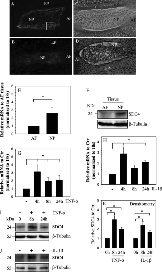
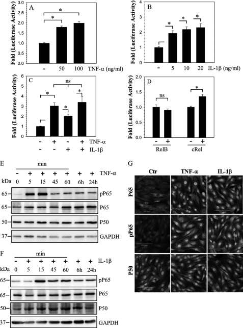
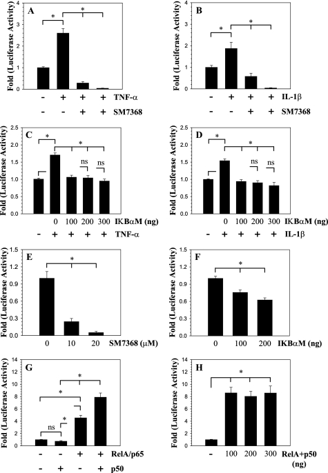
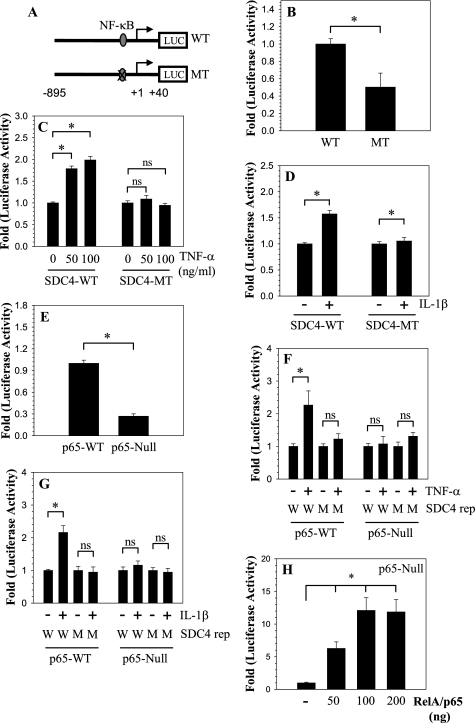
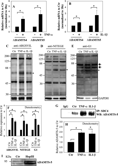
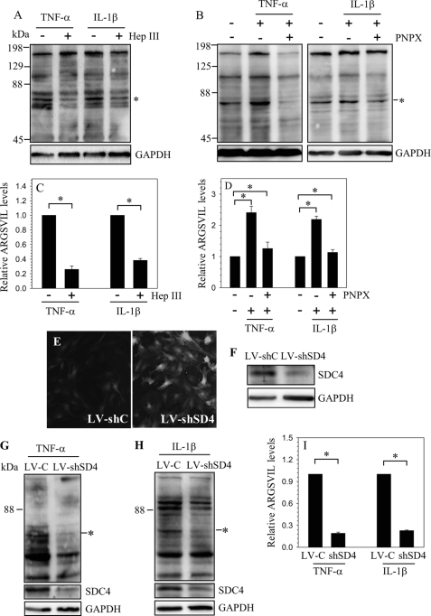
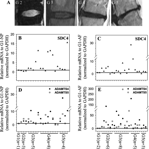
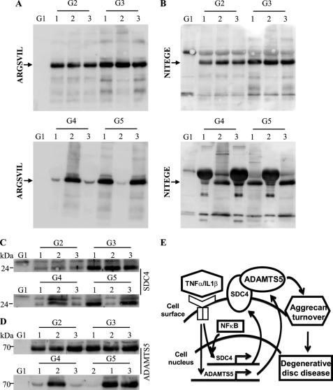
Elevated levels of TNF-α, IL-1β and a resultant increase in ADAMTS (a disintegrin-like and metalloprotease with thrombospondin type I motifs) expression is seen during disc degeneration. However, if these pro-inflammatory cytokines control ADAMTS activity is not definitively known. The goal of the investigation was to study if TNF-α and IL-1β regulate syndecan-4 (SDC4) expression, and if SDC4 was responsible for promoting aggrecan degradation through controlling ADAMTS activity in nucleus pulposus cells of the intervertebral disc. Cytokine treatment increased SDC4 expression and promoter activity. Use of inhibitor, SM7368 and co-transfections with IκBα, RelA/p50 showed that NF-κΒ regulated both basal and cytokine-dependent SDC4 transcription. SDC4 promoter harboring RelA binding site mutation was unresponsive to the cytokines. Moreover, cytokines failed to increase SDC4 promoter activity in RelA-null cells. Cytokines increased ADAMTS-4/5 expression and aggrecan degradation and promoted SDC4 interaction with ADAMTS-5. Treatment with heparinase-III and p-nitrophenyl-β-D-xylopyranoside (PNPX), an inhibitor of heparan sulfate synthesis and transfection with SDC4-shRNA partially blocked cytokine mediated aggrecan degradation. Analysis of human tissues showed increased aggrecan degradation with a concomitant increase in SDC4 and ADAMTS-5 protein expression with severity of disc disease. Likewise, SDC4, TNF-α, IL-1β, ADAMTS-4, and ADAMTS-5 mRNA expression increased in degenerate tissues. We conclude that in nucleus pulposus, TNF-α and IL-1β regulate SDC4 expression, which plays a key role in pathogenesis of degenerative disc disease by promoting aggrecan degradation by ADAMTS-5.


Figures

FIGURE 1.
FIGURE 1.
Expression and cytokine dependence of SDC4 in NP cells. Sagittal sections of the intervertebral disc of a mature (A, C) and a neonatal rat (B, D). Sections were treated with SDC4 antibody (A, B), or counterstained with hemotoxylin, eosin, and alcian blue (C and D). Cell surface staining of SDC4 was evident in NP cells in mature disc (box in A), SDC4 was expressed at a higher level in NP of the compared with the AF. Mag. × 4–10). E, real-time RT-PCR analysis shows significantly higher expression of SDC4 in NP than AF tissue. F, Western blot analysis of SDC4 expression in AF and NP tissue. Note, the expression of the 24–26 kDa SDC4 band, higher expression of SDC4 in NP compared with AF tissue. G and H, real-time RT-PCR analysis of SDC4 expression by nucleus pulposus cells treated with TNF-α (G) and IL-1β (H) up to 24 h. There was increased expression at 4 h which continued until 24 h. I and J, Western blot analysis showing increased expression of SDC4 by NP cells following TNF-α (I) and IL-1β (J) treatment. K, densitometric analysis of three blots each for experiment described in I and J above. β-Tubulin was used as a loading control and to calculate relative expression levels. Cytokine treatment significantly increased SDC4 expression. Values shown are mean ± S.E. of three independent experiments; *, p < 0.05.
FIGURE 2.
FIGURE 2.
Modulation of SDC4 promoter activity and NF-κB signaling by cytokines in NP cells. Treatment with both TNF-α (A) and IL-1β (B) induces activity of SDC4 promoter in NP cells. C, a combined treatment of TNF-α and IL-1β shows no additive or synergistic effect on induction of SDC4 promoter activity over individual cytokine treatments. D, NP cells were co-transfected with RelB or cRel and SDC4 promoter activity measured. Only cRel has a small inductive effect on SDC4 promoter activity. Values shown are mean ± S.E., of three independent experiments; *, p < 0.05. E and F, Western blot analysis of NF-κB signaling proteins, p65 and p50 following treatment of NP cells with TNF-α (E) and IL-1β (F). Cytokine treatment induced phosphorylation of p65 within first 15 min and levels remained elevated after 24 h. No appreciable change in expression of p65 and p50 was seen. G, immunofluorescent analysis of NP cells treated with TNF-α and IL-1β. Cells showed increased nuclear localization of both phospho-p65 and p65 whereas p50 expression and localization did not change.
FIGURE 3.
FIGURE 3.
Effect of NF-κB on SDC4 promoter activity in NP cells. A and B, SDC4 promoter activity was measured following TNF-α (A) and IL-1β (B) treatment of NP cells with or without NF-κB signaling inhibitor SM7368. Inhibitor treatment completely abolished cytokine mediate promoter induction. C and D, cells were co-transfected with IκBαM and promoter activity was measured following TNF-α (C) and IL-1β (D) treatment. Inhibition of NF-κB signaling resulted in significant blocking of cytokine dependent induction in SDC4 promoter activity. E, treatment of NP cells with inhibitor SM7368 or F, co-transfection with IKBαM, in absence of exogenous cytokines resulted in suppression of basal SDC4 promoter activity. G, NP cells were co-transfected with RelA/p65 and/or p50 and SDC4 promoter activity measured. Co-transfection with p65 alone but not p50 resulted in increased promoter activity. When p65 and p50 are added together they elicited a higher induction in activity than p65 alone. H, note the significant induction in SDC4 reporter activity even when transfected with 100 ng of total RelA and p50 plasmids. No further induction in activity was seen when plasmid concentration was increased till 300 ng. Values shown are mean ± S.E. from three independent experiments, *, p < 0.05.
FIGURE 4.
FIGURE 4.
NF-κB regulation of SDC4 expression. A, schematic of different SDC4 reporter constructs (WT: wild type; MT: mutant RelA). B, cells were transfected with WT and MT SDC4 reporter constructs and basal activity measured. Mutant promoter exhibits significantly lower basal activity than the wild type promoter. C and D, cells transfected with WT and MT SDC4 reporter constructs were treated with C, TNF-α and D, IL-1β. Only the WT SDC4 promoter fragment with elicited an increase in activity. E, SDC4 promoter activity in MEFs isolated from RelA WT and null mice. There was a 70% decrease in basal SDC4 reporter activity in the RelA-null cells. F and G, RelA WT and null cells transfected with WT and MT SDC4 reporter constructs and treated with F, TNF-α and G, IL-1β. Wild type cells show increase in only WT reporter activity. While null cells do not show increase either WT or MT promoter. H, RelA-null cells were transfected with plasmid expressing RelA and SDC4 promoter activity was measured. A dose-dependent increase in SDC4 reporter activity was seen, at a concentration higher than 200 ng of RelA no further increase in activity was seen. Values shown are mean ± S.E. from three independent experiments, *, p < 0.05.
FIGURE 5.
FIGURE 5.
Regulation of ADAMTS expression and activity by TNF-α and IL-1β. Real-time RT-PCR analysis of cells treated with A, TNF-α and B, IL-1β show increase in expression of ADAMTS-4 and ADAMTS-5. C–E, Western blot analysis to measure aggrecan degradation using neoepitope antibodies C, anti-ARGSVIL D, anti-NITEGE E, anti-G1. A prominent increase in degradation products corresponding to particular epitopes (marked by asterisk and arrows). F, densitometric analysis of three blots each for experiment described in C, D, and E above. GAPDH was used as a loading control and to calculate relative expression. Increase in degradation products corresponding to epitopes marked by asterisk in C–E was seen by the cytokine treatment. G, immunoprecipitation (IP) of SDC4 from control and cytokine-treated cells followed by a Western blot using ADAMTS-5 antibody shows increased association between SDC4 and ADAMTS-5 in treated cells. IP using an isotype IgG in place of SDC4 antibody showed absence of ADAMTS-5 co-precipitation. H, densitometric analysis of three blots for experiment described in G above shows increased co-precipitation of SDC4 and ADAMTS-5 in cytokine-treated NP cells. I, treatment of NP cells with heparinase (Hep) III resulted in increased shedding of pro-ADAMTS-5 in the medium. Values shown in quantitative experiments are mean ± S.E. from three independent experiments, *, p < 0.05.
FIGURE 6.
FIGURE 6.
Regulation of ADAMTS activity by SDC4. Western blot analysis of aggrecan neoepitope (ARGSVIL) in cells treated with cytokines with or without A, Heparinase (Hep) III or B, PNPX. A prominent decrease in aggrecan neoepitope formation is seen when cells are treated with either heparinase or PNPX. C and D, densitometric analysis of three blots each for experiment described in A and B, respectively. GAPDH was used as a loading control and to calculate relative expression. A significant decrease in aggrecan neoepitope formation is seen in cells treated with either heparinase or PNPX. E, immunofluorescent detection of GFP in NP cells transduced with lentivirus co-expressing GFP and control ShRNA (LV-shC) and lentivirus co-expressing GFP and SDC4 shRNA (LV-shSD4) shows high transduction efficiency. Magnification ×20. F, Western blot analysis of cells infected with LV-shC and LV-shSD4. The expression of SDC4 was suppressed by SDC4 shRNA compared with cells transduced with control lentivirus. G and H, Western blot analysis of aggrecan neoepitope (ARGSVIL) in LV-shC and LV-shSD4 transduced cells treated with G, TNF-α and H, IL-1β. I, densitometric analysis of three blots each for experiment described in G and H. Note that aggrecan neoepitope generation is significantly reduced following cytokine treatment in SDC4-silenced cells compared with controls. Values shown in quantitative experiments are mean ± S.E. from three independent experiments, *, p < 0.05.
FIGURE 7.
FIGURE 7.
SDC4 and ADAMTS-4/5 mRNA expression in human degenerate NP and AF tissues. A, representative MRI images of patients with degenerative disc disease. (From left, Grade II,III,IV,V). B–E, real-time RT-PCR analysis of SDC4 (B and C) and ADAMTS4/5 (D and E) mRNA expression in multiple human NP (B and D) and AF (C and E) tissue samples. Samples from multiple different patients were used for each degenerative grade. Compared with normal control, with increasing disc degeneration there is a relative increase in the expression of SDC4 and ADAMTS mRNA. Mean expression of normal controls (c) was set at 1.0 and shown by a horizontal line.
FIGURE 8.
FIGURE 8.
Increased aggrecan degradation is correlated with SDC4 and ADAMTS-5 protein levels in degenerate human NP tissues. A and B, Western blot analysis of ADAMTS generated aggrecan neoepitopes in human degenerate NP tissue samples using A, anti-ARGSVIL and B, anti-NITEGE antibodies. Both ARGSVIL and NITEGE neoepitope levels are significantly higher in all the degenerate NP samples compared with control tissue. Control sample showed undetectable levels of these aggrecan degradation products. Samples from three different patients were used for each degenerative grade. C, SDC4 and D, ADAMTS-5 expression levels in corresponding human degenerate NP samples. Degenerate samples that exhibit high level of aggrecan neoepitope accumulation also showed a high expression of both SDC4 and ADAMTS-5. Expression of SDC4 and ADAMTS-5 was very low in control sample. E, a proposed model of relationship between inflammatory cytokines (TNF-α, IL-1β), SDC4, and ADAMTS-5 in NP cells. During degeneration high levels of inflammatory cytokines TNF-α and IL-1β transcriptionally induce SDC4 expression through NF-κB signaling pathway. Increased level of SDC4 binds and anchors ADAMTS-5 on cell surface for processing and results in its activation. Active ADAMTS-5 cleaves aggrecan matrix resulting in loss of water binding capacity and disc height.

Similar articles

Cited by

References

    1. Feng H., Danfelter M., Stromqvist B., Heinegard D. (2006) J Bone Joint Surg Am 88, Suppl. 2, 25–29 - PubMed
    1. Setton L.A., Chen J. (2006) J. Bone Joint Surg. Am. 88, Suppl. 2, 52–57 - PubMed
    1. Ng L, Grodzinsky A.J., Patwari P., Sandy J., Plaas A., Ortiz C. (2003) J. Struct. Biol. 143, 242–257 - PubMed
    1. Roberts S., Evans H., Trivedi J., Menage J. (2006) J. Bone Joint Surg. Am. 88, Suppl. 2, 10–14 - PubMed
    1. Miljkovic D., Trajkovic V. (2004) Cytokine Growth Factor Rev. 15, 21–32 - PubMed

Publication types

MeSH terms

Substances