. 2012 Jan 1;21(1):76-84.
doi: 10.1093/hmg/ddr439. Epub 2011 Sep 23.

Protein interacting with C kinase (PICK1) is a suppressor of spinocerebellar ataxia 3-associated neurodegeneration in Drosophila

Affiliations

Protein interacting with C kinase (PICK1) is a suppressor of spinocerebellar ataxia 3-associated neurodegeneration in Drosophila

Leeanne McGurk et al. Hum Mol Genet. .

Abstract

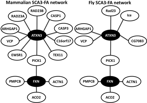
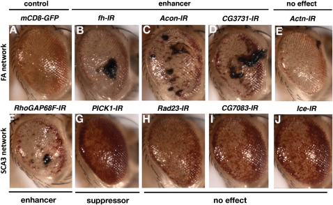
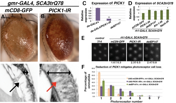
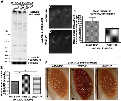
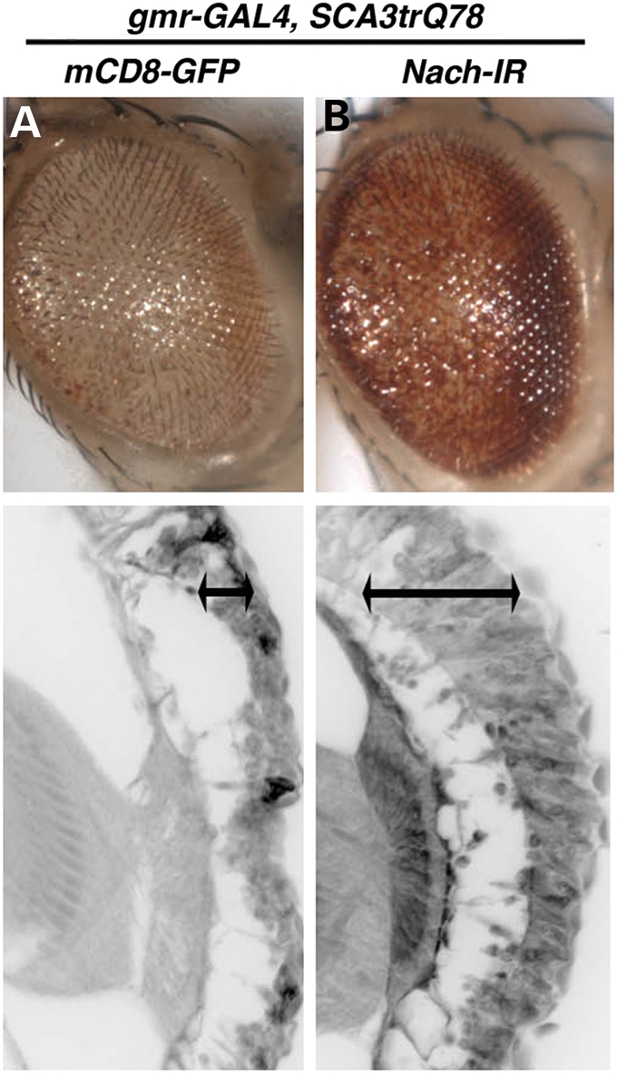
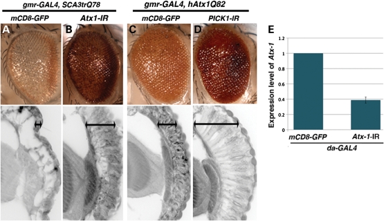
Spinocerebellar ataxia 3 (SCA3) is the most common autosomal dominant ataxia. The disease is caused by an expansion of a CAG-trinucelotide repeat region within the coding sequence of the ATXN3 gene, and this results in an expanded polyglutamine (polyQ) tract within the Ataxin-3 protein. The polyQ expansion leads to neuronal dysfunction and cell death. Here, we tested the ability of a number of proteins that interact with Ataxin-3 to modulate SCA3 pathogenicity using Drosophila. Of 10 candidates, we found four novel enhancers and one suppressor. The suppressor, PICK1 (Protein interacting with C kinase 1), is a transport protein that regulates the trafficking of ion channel subunits involved in calcium homeostasis to and from the plasma membrane. In line with calcium homeostasis being a potential pathway mis-regulated in SCA3, we also found that down-regulation of Nach, an acid sensing ion channel, mitigates SCA3 pathogenesis in flies. Modulation of PICK1 could be targeted in other neurodegenerative diseases, as the toxicity of SCA1 and tau was also suppressed when PICK1 was down-regulated. These findings indicate that interaction proteins may define a rich source of modifier pathways to target in disease situations.


Figures

Figure 1.
Figure 1.
The SCA3-FA network. The ataxia interactome was used to predict protein interactors of Ataxin-3 in Drosophila (14). Out of the 10 direct interactors of Ataxin-3, 8 have a clear sequence counterpart in Drosophila. Ataxin-3 is one node away from frataxin, the protein responsible for FA. Due to the histopathological similarities between these two diseases, we chose to include the FA-network in our analysis.
Figure 2.
Figure 2.
SCA3 candidate modifiers. (A) Expression of strong SCA3trQ78 causes degeneration of the external fly eye. Genotype: w; gmr-GAL4, UAS-SCA3trQ78/UAS-mCD8-GFP. (B–E) Genes of the frataxin network. Genotypes: w; gmr-GAL4, UAS-SCA3trQ78 in trans to the alleles indicated. (B–D) Genes of the frataxin network that enhance the SCA3trQ78 degenerate eye phenotype. (E) Reduction of Actn, a frataxin network protein, has no effect on the SCA3trQ78 degenerate eye phenotype. (F–J) Genes of the SCA3 network. Genotypes: w; gmr-GAL4, UAS-SCA3trQ78 in trans to the alleles indicated. (F) RhoGAP68F when knocked-down enhances the SCA3trQ78 degenerate eye phenotype. (G) Reduction in PICK1 suppresses the SCA3trQ78 eye phenotype. (H–J) Genes of the SCA3 network, when knocked-down, that have no effect on the SCA3trQ78 degenerate eye phenotype.
Figure 3.
Figure 3.
Reduction in PICK1 is neuroprotective in SCA3. (A) Expression of SCA3trQ78 causes external and internal degeneration, with pigment loss and disintegration of retinal structure. Genotype w; gmr-GAL4, UAS-SCA3trQ78/UAS-mCD8-GFP. Black arrow indicates loss of tissue, and double-headed arrow indicates retinal tissue width. (B) Knockdown of PICK1 gene expression suppresses the degeneration of the SCA3trQ78 external eye and deterioration of the retinal structure. Genotype w; gmr-GAL4, UAS-SCA3trQ78/UAS-PICK1-IR. The red arrow indicates preservation of retinal tissue, and double-headed arrow indicates retinal tissue width. (C) Global expression of UAS-PICK1-IR with the daughterless-GAL4 (da-GAL4) driver causes a consistent knock-down of PICK1 expression compared with the control, mean ± SEM is shown, *P< 0.001. (D) The suppression of the SCA3trQ78 degenerate eye phenotype is not due to a reduction in SCA3 transgene expression. One-way analysis of variance (ANOVA) was performed followed by a Tukey's post-test, *P< 0.05, mean ± SEM. (E) Photoreceptor neural degeneration is suppressed by PICK1 reduction. Photoreceptor neurons were counted in eyes of flies by optical neutralization. Representative images of each genotype are shown and the mean ± SEM number of ommatidia is indicated below each image. An aged-matched wild-type control shows the expected non-degenerated ommatidial structure with seven photoreceptors. Data are 10–20 ommatidia of at least 10 flies per genotype, repeated three independent times. (F) The distribution of ommatidia in female adults at d21. Expression of SCA3trQ78 in the photoreceptor neurons with the rhodopsin-1-GAL4 (rh1-GAL4) driver results in a severe loss in the number of photoreceptor neurons at day 21. Reduction in PICK1, either by the co-expression of the UAS-PICK1 RNAi transgene or by heterozygous loss of endogenous PICK1, suppresses the photoreceptor loss at day 21, *P< 0.001, mean ± SEM.
Figure 4.
Figure 4.
Reduction in PICK1 expression in the SCA3trQ78 eye leads to less insoluble SCA3 protein. (A) Western blot detecting SCA3trQ78 with anti-HA shows that reduction in PICK1 expression decreases the amount of insoluble SCA3trQ78 protein, present in the stacking gel, and increases the amount of soluble SCA3trQ78 protein. Genotype w; rh1-GAL4, UAS-SCA3trQ78 in trans to indicated alleles. (B) Quantification of the mean ± SEM of six independent repeats showing the ratio of soluble protein relative to the control strain (normalized to tubulin). One-way ANOVA followed by a Tukey's test, *P< 0.05. (C and D) Horizontal cryosections of flies at d3 stained with anti-HA to detect SCA3trQ78. (C) Control flies showing the accumulation of insoluble SCA3trQ78. Genotype w; UAS-mCD8-GFP /+; gmr-GAL4, UAS-SCA3trQ78+. (D) Flies with reduced PICK1 expression show a slight reduction in the accumulation of the SCA3trQ78 protein. Genotype w; gmr-GAL4, UAS-SCA3trQ78/UAS-PICK1-IR (E) SCA3trQ78 inclusions were quantified in cryosections from animals expressing SCA3trQ78 compared with those expressing the disease protein but now with reduced PICK1 levels, mean ± SEM. (F) Flies expressing hTau.R406W in the eye show a degenerate, reduced eye. Reduction in PICK1 suppresses the small eye. The PICK1 deletion line delEP147 did not suppress the small eye phenotype as robustly as the RNAi line, although the RNAi line reduces PICK1 expression to a much greater effect than the deletion. Genotype: w, gmr-GAL4; UAS-htau.R406W in trans to indicated alleles. Double-headed arrow indicates length of fly eye.
Figure 5.
Figure 5.
Reduction in Nach, an ASIC, mitigates SCA3 pathogenesis. (A) Control strain showing degeneration of the external eye and of the retina. Genotype: w; gmr-GAL4, UAS-SCA3trQ78/UAS-mCD8-GFP. Double-headed arrow indicates retinal tissue width. (B) Co-expression of an RNAi transgene to Nach, an ASIC, suppresses the external internal degeneration induced by SCA3trQ78. Genotype: w; gmr-GAL4, UAS-SCA3trQ78/UAS-Nach-IR. Double-headed arrow indicates retinal tissue width.
Figure 6.
Figure 6.
SCA1 shares common pathogenic pathways to SCA3. (A) Control strain shows degeneration of the external eye and of the retina. Genotype: w; gmr-GAL4, UAS-SCA3trQ78/UAS-mCD8-GFP. (B) Co-expression of an RNAi transgene directed to Atx-1 suppresses the SCA3 degeneration. w; gmr-GAL4, UAS-SCA3trQ78/UAS-Atx-1-IR. (C and D) Reduction in PICK1 gene expression suppresses hAtx1Q82 degeneration. (C) Expression of the human Ataxin-1 protein with an expansion of Q82 causes degeneration of the external eye and of the retina. Genotype: w-; UAS-mCD8-GFP/+; gmr-GAL4 (YHIII), UAS-hAtx1-Q82. (D) Co-expression of the RNAi transgene directed to PICK1 suppresses the SCA1-associated degeneration. Genotype: w-; gmr-GAL4, UAS-Atx1 Q82/UAS-PICK1-IR. Double-headed arrow indicates retinal tissue width. (E) Global expression, with the da-GAL4 driver, of the RNAi transgene directed to Atx-1 consistently reduces endogenous Atx-1 gene levels (mean ± SEM, P < 0.03).

References

    1. Davies S.W., Turmaine M., Cozens B.A., DiFiglia M., Sharp A.H., Ross C.A., Scherzinger E., Wanker E.E., Mangiarini L., Bates G.P. Formation of neuronal intranuclear inclusions underlies the neurological dysfunction in mice transgenic for the HD mutation. Cell. 1997;90:537–548. - PubMed
    1. DiFiglia M., Sapp E., Chase K.O., Davies S.W., Bates G.P., Vonsattel J.P., Aronin N. Aggregation of huntingtin in neuronal intranuclear inclusions and dystrophic neurites in brain. Science. 1997;277:1990–1993. - PubMed
    1. Paulson H.L., Perez M.K., Trottier Y., Trojanowski J.Q., Subramony S.H., Das S.S., Vig P., Mandel J.L., Fischbeck K.H., Pittman R.N. Intranuclear inclusions of expanded polyglutamine protein in spinocerebellar ataxia type 3. Neuron. 1997;19:333–344. - PubMed
    1. Poirier M.A., Li H., Macosko J., Cai S., Amzel M., Ross C.A. Huntingtin spheroids and protofibrils as precursors in polyglutamine fibrilization. J Biol. Chem. 2002;277:41032–41037. - PubMed
    1. Dawson T.M., Dawson V.L. Molecular pathways of neurodegeneration in Parkinson's disease. Science. 2003;302:819–822. - PubMed

Publication types

MeSH terms