. 2011 Oct 11;108(41):16927-31.
doi: 10.1073/pnas.1112047108. Epub 2011 Sep 26.

Nuclear receptor liver receptor homologue 1 (LRH-1) regulates pancreatic cancer cell growth and proliferation

Affiliations

Nuclear receptor liver receptor homologue 1 (LRH-1) regulates pancreatic cancer cell growth and proliferation

Cindy Benod et al. Proc Natl Acad Sci U S A. .

Abstract

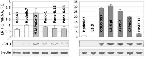
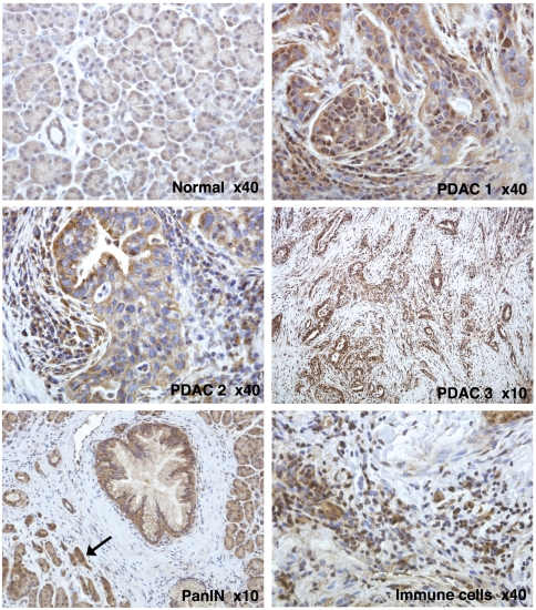
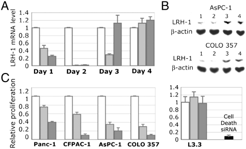
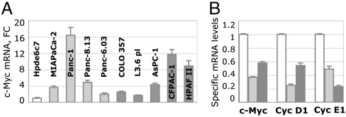
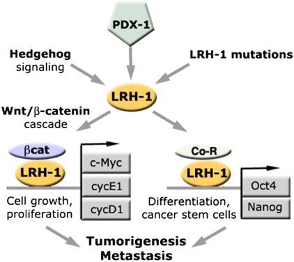
An essential regulator of gene transcription, nuclear receptor liver receptor homologue 1 (LRH-1) controls cell differentiation in the developing pancreas and maintains cholesterol homeostasis in adults. Recent genome-wide association studies linked mutations in the LRH-1 gene and its up-stream regulatory regions to development of pancreatic cancer. In this work, we show that LRH-1 transcription is activated up to 30-fold in human pancreatic cancer cells compared to normal pancreatic ductal epithelium. This activation correlates with markedly increased LRH-1 protein expression in human pancreatic ductal adenocarcinomas in vivo. Selective blocking of LRH-1 by receptor specific siRNA significantly inhibits pancreatic cancer cell proliferation in vitro. The inhibition is tracked in part to the attenuation of the receptor's transcriptional targets controlling cell growth, proliferation, and differentiation. Previously, LRH-1 was shown to contribute to formation of intestinal tumors. This study demonstrates the critical involvement of LRH-1 in development and progression of pancreatic cancer, suggesting the LRH-1 receptor as a plausible therapeutic target for treatment of pancreatic ductal adenocarcinomas.


Conflict of interest statement

The authors declare no conflict of interest.

Figures

Fig. 1.
Fig. 1.
Expression of LRH-1 in pancreatic cancer cells. Levels of LRH-1 mRNA in pancreatic cancer cells originated from primary and metastatic PDACs (Left, Right, in light and dark gray) are shown relative to control (nonneoplastic pancreatic ductal epithelial cells Hpde6c7, in white); note 10-fold scale change in the right panel. (Left) For comparison, the levels of LRH-1 mRNA in hepatocarcinoma cells HepG2 are shown in white. Standard deviations are drawn as black lines. Western blots at the bottom of the panels show normalized expression of LRH-1 protein in the corresponding cells (imaged by enhanced chemiluminescence, 1-min exposure). The expression levels of LRH-1 are normalized to those of β-actin (detected after 10-s exposure).
Fig. 2.
Fig. 2.
Differential expression of LRH-1 in normal and cancerous pancreas. Shown are IHC images of human normal (Normal) and neoplastic pancreas (PDAC 1–3). The samples were treated with primary antibody HPA005455 (Sigma-Aldrich), stained with ImmPACT diaminobenzidine (Vector Labs) and counterstained with hematoxylin; the original magnification (× 40 or × 10) is specified. Weak cytoplasmic and weak-to-moderate punctate nuclear staining is observed for all components of normal exocrine pancreas. In neoplastic cells, elevated levels of LRH-1 are observed either in the nuclei (PDAC 1) or in the cytoplasm (PDAC 2) or both. Whereas the fibroblasts surrounding neoplastic lesions do not show significant LRH-1 specific staining (PDAC 3), many infiltrating immune cells (indicated) are positively stained for LRH-1. Heightened levels of LRH-1 are also observed in PanIN lesions (indicated) and in the acinar and ductal cells affected by pancreatitis (indicated by arrow).
Fig. 3.
Fig. 3.
Blocking LRH-1 function by siRNA specific to the receptor. (A) Inhibition of LRH-1 transcription in AsPC-1 cells. Cells were analyzed by qPCR for relative levels of LRH-1 mRNA at days 1–4 following transfections. Control in white corresponds to cells treated with irrelevant siRNA (see Materials and Methods); light- and dark-gray bars indicate mRNA in cells transfected with two LRH-1 specific siRNAs. (B) Western blot analyses of AsPC-1 and COLO 357 cells transfected with anti-LRH-1 siRNAs. Lanes: 1 and 2, cells treated with two LRH-1 specific siRNA; 3, cells transfected with control siRNA; 4, nontransfected cells. Data are shown for day 2 following the transfections. Expression of β-actin was used as an internal control. (C) Effect of LRH-1 knockdown on cancer cell proliferation. Proliferation of four pancreatic cancer cells expressing LRH-1 (indicated in the left panel) was compared following their transfections with two LRH-1 specific siRNAs (in light and dark gray). Data are shown for day 4 relative to the control (white). Proliferation of cells L3.3 that do not express LRH-1 was not affected by the anti-LRH-1 siRNAs (shown in the right panel relative to the control). The efficiency of transfections (> 90%) was optimized by monitoring proliferation of cells treated with AllStars Hs Cell Death Control siRNA (in black).
Fig. 4.
Fig. 4.
Effects of blocking LRH-1 on transcription of genes controlling cell proliferation. (A) Quantitative PCR data showing levels of C-Myc mRNA. Levels of C-Myc in primary and metastatic cells are shown as light- and dark-gray bars, relative to that found in nonneoplastic cells Hpde6c7. Standard deviations are drawn as black lines. (B) Inhibition of C-Myc, Cyc D1, and Cyc E1 gene transcription in AsPc-1 cells treated with anti-LRH-1 siRNA. Cells were analyzed by qPCR for the relative levels of mRNA corresponding to C-Myc, Cyc D1, and Cyc E1 (indicated). Controls in white correspond to cells treated with irrelevant siRNA; light- and dark-gray bars indicate the levels of corresponding mRNA in cells transfected with two LRH-1 specific siRNAs.
Fig. 5.
Fig. 5.
Possible mechanisms of involvement of nuclear receptor LRH-1 in development of pancreatic cancer. Indicated are major transcriptional targets of LRH-1 and regulatory pathways mediated by the receptor. The expression of LRH-1 (yellow symbol) is controlled by the pancreas-specific master transcription factor PDX-1 (indicated in green) and via hedgehog signaling pathway that are both reactivated in pancreatic cancer. Activating mutations in the LRH-1 gene and its regulatory regions might contribute to the receptor up-regulation. The activated receptor functions synergistically with β-catenin (shown in purple), activating multiple target genes regulating cell growth and proliferation (Cyc D1, Cyc E1, and C-Myc, indicated in gray) as well as cell differentiation (Oct4, Nanog, indicated in gray). The cumulative effect of the aberrant, LRH-1 mediated activation of these multiple targets in adult pancreas results in tumorigenesis and metastasis.

References

    1. Thayer SP, et al. Hedgehog is an early and late mediator of pancreatic cancer tumorigenesis. Nature. 2003;425:851–856. - PMC - PubMed
    1. Pasca di Magliano M, et al. Common activation of canonical Wnt signaling in pancreatic adenocarcinoma. PLoS One. 2007;2:e1155. - PMC - PubMed
    1. Fayard E, Auwerx J, Schoonjans K. LRH-1: An orphan nuclear receptor involved in development, metabolism and steroidogenesis. Trends Cell Biol. 2004;14:250–260. - PubMed
    1. Fayard E, Schoonjans K, Annicotte J-S, Auwerx J. Liver receptor homolog 1 controls the expression of carboxyl ester lipase. J Biol Chem. 2003;278:35725–35731. - PubMed
    1. Kim JW, Peng N, Rainey WE, Carr BR, Attia GR. Liver receptor homolog-1 regulates the expression of steroidogenic acute regulatory protein in human granulosa cells. J Clin Endocrinol Metab. 2004;89:3042–3047. - PubMed

Publication types

MeSH terms

Substances