. 2011 Nov 15;108(46):18649-54.
doi: 10.1073/pnas.1113170108. Epub 2011 Sep 26.

UBLCP1 is a 26S proteasome phosphatase that regulates nuclear proteasome activity

Affiliations

UBLCP1 is a 26S proteasome phosphatase that regulates nuclear proteasome activity

Xing Guo et al. Proc Natl Acad Sci U S A. .

Abstract

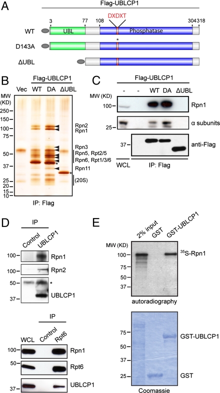
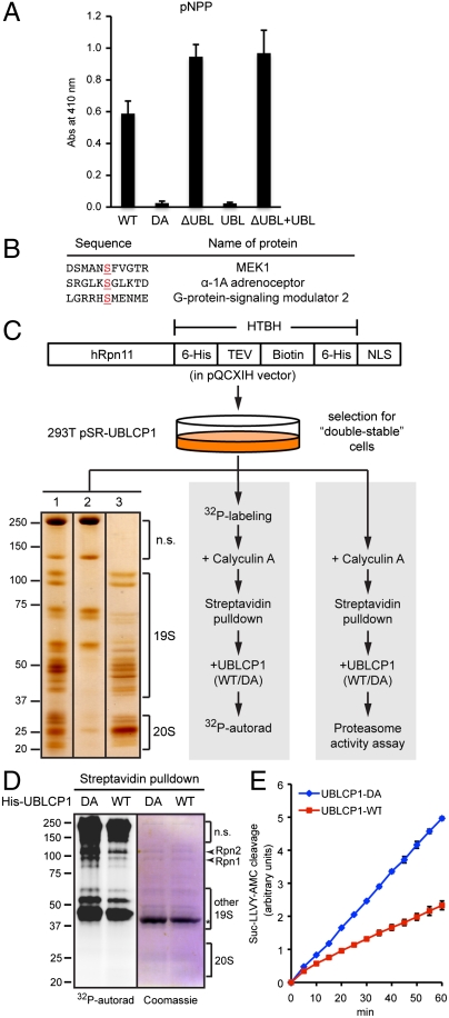
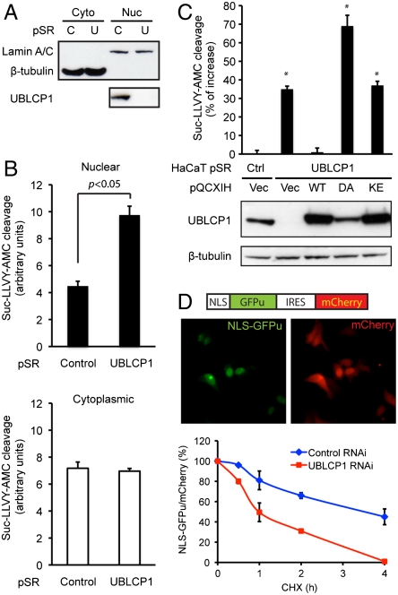
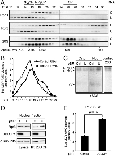
Protein degradation by the 26S proteasome is a fundamental process involved in a broad range of cellular activities, yet how proteasome activity is regulated remains poorly understood. We report here that ubiquitin-like domain-containing C-terminal domain phosphatase 1 (UBLCP1) is a 26S proteasome phosphatase that regulates nuclear proteasome activity. UBLCP1 directly interacts with the proteasome via its UBL domain and is exclusively localized in the nucleus. UBLCP1 dephosphorylates the 26S proteasome and inhibits proteasome activity in vitro. Knockdown of UBLCP1 in cells promotes 26S proteasome assembly and selectively enhances nuclear proteasome activity. Our results describe the first identified proteasome-specific phosphatase and uncover a unique mechanism for phosphoregulation of the proteasome.


Conflict of interest statement

The authors declare no conflict of interest.

Figures

Fig. 1.
Fig. 1.
UBLCP1 binds the 26S proteasome. (A) A schematic representation of UBLCP1 used for generating 293T stable lines used in B. All constructs contain a N-terminal Flag tag (gray oval). (B) UBLCP1-interacting proteins visualized by silver staining. Proteasome subunits identified by MS are indicated by arrowheads. 20S subunits were verified by Western blot. Asterisks indicate the bait Flag-UBLCP1 proteins. (C) Coimmunoprecipitation of Flag-UBLCP1 with RP (Rpn1) and CP (α subunits). (D) Binding between endogenous UBLCP1 and the proteasome. Preimmune rabbit serum or normal mouse IgG was used as controls for UBLCP1 and Rpt6 IP, respectively. Asterisk indicates the heavy chain of rabbit IgG. (E) In vitro binding between UBLCP1 and Rpn1.
Fig. 2.
Fig. 2.
Functional importance of the UBL domain. (A) Structural comparison of ubiquitin (PDB ID code 1UBQ) and the UBL domains of Drosophila UBLCP1 (PDB ID code 3SHQ), yeast Dsk2 (PDB ID code 2BWF), and human Rad23A (PDB ID code 2WYQ). The side chains of the KLL motif (Lys46/Leu47/Leu48) in DmUBLCP1 and its counterparts in the other structures are shown. (B) 293T cells were transfected with Flag-UBLCP1 constructs (G10E, L46A, K44E, and K44A/L45A/L46A) and were subjected to coimmunoprecipitation assays. (C) Immunostaining of endogenous UBLCP1 and Rpt6 in HaCaT cells (human keratinocytes). Scale bar = 10 μm. (D) Localization of GFP-tagged UBLCP1 and mutants in HeLa cells. Nuclear contours are depicted with white dotted lines based on DAPI images. Scale bar = 10 μm.
Fig. 3.
Fig. 3.
UBLCP1 dephosphorylates the proteasome in vitro. (A) pNPP assay with the indicated UBLCP1 proteins. Data are shown as average ± SD. (B) Phospho-peptides significantly dephosphorylated by UBLCP1 in vitro, with the phospho-serine residues underlined. This phospho-peptide array (Millipore) does not include any proteasome peptides. (C) 26S proteasome isolation and analysis. 293T double-stable line was used for high-purity proteasome preparation (Left), 32P-labeling and in vitro phosphatase assay (Middle), or in vitro proteasome activity assay (Right). (Left) A representative silver-staining image of streptavidin-immobilized proteasomes (lane 1), purified proteasomes after tobacco etch virus (TEV) protease treatment (lane 3), and nonspecific (n.s.) proteins remaining on the beads (lane 2). See Materials and Methods for details. (D) Metabolically radio-labeled, affinity-purified nuclear proteasomes were treated in vitro with purified UBLCP1 (shown by an asterisk). Proteasome subunits were visualized by Coomassie blue staining and 32P-autoradiography. (E) Peptidase activity assay of the 26S proteasome purified and treated as in D. AMC fluorescence is represented as the average ± SD.
Fig. 4.
Fig. 4.
UBLCP1 knockdown enhances nuclear proteasome activity. (A) Cytoplasmic (Cyto) and nuclear (Nuc) fractionation of ZR751 breast cancer cells stably expressing either control (C) or UBLCP1-specific (U) shRNA built in the pSuperRetro vector (pSR). Lamin A/C and β-tubulin were used as loading controls for nuclear and cytoplasmic fractions, respectively. (B) Proteasome activity assay using the same extracts as in A. Normalized AMC fluorescence is represented as the average ± SD. (C) UBLCP1 rescue experiments with HaCaT double-stable lines. UBLCP1 expression levels are determined by Western blotting. Nuclear proteasome activity was measured by in-well assays. Data are presented as the percent increase in activity versus control. *, p < 0.05. (D, Top) A schematic of the NLS-GFPu reporter in an IRES-mCherry backbone (26) and localization of the fluorescent proteins in 293T cells. (Bottom) GFPu/mCherry ratio during cycloheximide (CHX) treatment time course, with the starting value at time 0 being 100%. Data were analyzed from three independent experiments.
Fig. 5.
Fig. 5.
UBLCP1 regulates RP-CP association. (A) Size-exclusion chromatography analysis of proteasome complexes from 293T cells stably expressing control or UBLCP1 shRNA. Proteasome subunits within each fraction were monitored by Western blot. The different forms of proteasome are shown (Top) based on their subunit composition and their expected sizes (Bottom). (B) Proteasome activity in gel-filtration fractions from A after normalization against the protein content within each fraction. (C) Gel overlay assay. Equal amounts of cytosolic and nuclear extracts from HaCaT pSuperRetro stable lines were separated on a 4.5% native gel. U, UBLCP1 shRNA. Purified 26S proteasome (1 μg; see Fig. 3C) was included as a positive control. The gel was then overlaid with proteasome assay buffer containing Suc-LLVY-AMC and 0.02% SDS (to detect 20S activity). After incubation in the dark at 37 °C for 30 min, the gel was imaged under UV light. (D and E) Coimmunoprecipitation of nuclear CP and RP from HaCaT pSuperRetro stable lines. Anti-CP immunoprecipitates were divided and used for Western blot (D) and proteasome activity assay (E). AMC fluorescence was normalized against the amount of immunoprecipitated CP.

Comment in

Similar articles

Cited by

References

    1. Hershko A, Ciechanover A. The ubiquitin system. Annu Rev Biochem. 1998;67:425–479. - PubMed
    1. Bossy-Wetzel E, Schwarzenbacher R, Lipton SA. Molecular pathways to neurodegeneration. Nat Med. 2004;10(Suppl):S2–S9. - PubMed
    1. Hoeller D, Dikic I. Targeting the ubiquitin system in cancer therapy. Nature. 2009;458:438–444. - PubMed
    1. Bingol B, Sheng M. Deconstruction for reconstruction: The role of proteolysis in neural plasticity and disease. Neuron. 2011;69:22–32. - PubMed
    1. Finley D. Recognition and processing of ubiquitin-protein conjugates by the proteasome. Annu Rev Biochem. 2009;78:477–513. - PMC - PubMed

Publication types

MeSH terms

Associated data