

Transmembrane segments of nascent polytopic membrane proteins control cytosol/ER targeting during membrane integration
- PMID: 21949411
- PMCID: PMC3187712
- DOI: 10.1083/jcb.201103117
Transmembrane segments of nascent polytopic membrane proteins control cytosol/ER targeting during membrane integration
Abstract
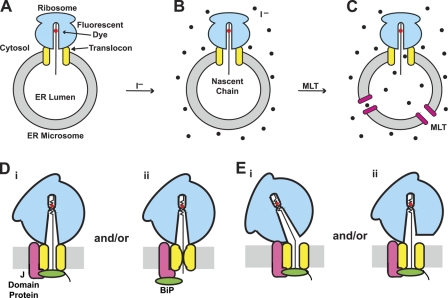
During cotranslational integration of a eukaryotic multispanning polytopic membrane protein (PMP), its hydrophilic loops are alternately directed to opposite sides of the ER membrane. Exposure of fluorescently labeled nascent PMP to the cytosol or ER lumen was detected by collisional quenching of its fluorescence by iodide ions localized in the cytosol or lumen. PMP loop exposure to the cytosol or lumen was controlled by structural rearrangements in the ribosome, translocon, and associated proteins that occurred soon after a nascent chain transmembrane segment (TMS) entered the ribosomal tunnel. Each successive TMS, although varying in length, sequence, hydrophobicity, and orientation, reversed the structural changes elicited by its predecessor, irrespective of loop size. Fluorescence lifetime data revealed that TMSs occupied a more nonpolar environment than secretory proteins inside the aqueous ribosome tunnel, which suggests that TMS recognition by the ribosome involves hydrophobic interactions. Importantly, the TMS-triggered structural rearrangements that cycle nascent chain exposure between cytosolic and lumenal occur without compromising the permeability barrier of the ER membrane.
Figures


Similar articles
-
Polytopic membrane protein folding at L17 in the ribosome tunnel initiates cyclical changes at the translocon.J Cell Biol. 2011 Oct 3;195(1):55-70. doi: 10.1083/jcb.201103118. Epub 2011 Sep 26. J Cell Biol. 2011. PMID: 21949410 Free PMC article.
-
The Ribosome-Sec61 Translocon Complex Forms a Cytosolically Restricted Environment for Early Polytopic Membrane Protein Folding.J Biol Chem. 2015 Nov 27;290(48):28944-52. doi: 10.1074/jbc.M115.672261. Epub 2015 Aug 7. J Biol Chem. 2015. PMID: 26254469 Free PMC article.
-
Both lumenal and cytosolic gating of the aqueous ER translocon pore are regulated from inside the ribosome during membrane protein integration.Cell. 1997 Jul 11;90(1):31-41. doi: 10.1016/s0092-8674(00)80311-6. Cell. 1997. PMID: 9230300
-
Functional ramifications of FRET-detected nascent chain folding far inside the membrane-bound ribosome.Biochem Soc Trans. 2004 Nov;32(Pt 5):668-72. doi: 10.1042/BST0320668. Biochem Soc Trans. 2004. PMID: 15493983 Review.
-
Biogenesis of CFTR and other polytopic membrane proteins: new roles for the ribosome-translocon complex.J Membr Biol. 2004 Dec;202(3):115-26. doi: 10.1007/s00232-004-0715-6. J Membr Biol. 2004. PMID: 15798900 Review.
Cited by
-
Cotranslational stabilization of Sec62/63 within the ER Sec61 translocon is controlled by distinct substrate-driven translocation events.Mol Cell. 2015 Apr 16;58(2):269-83. doi: 10.1016/j.molcel.2015.02.018. Epub 2015 Mar 19. Mol Cell. 2015. PMID: 25801167 Free PMC article.
-
Site-Directed Fluorescence Approaches for Dynamic Structural Biology of Membrane Peptides and Proteins.Front Mol Biosci. 2019 Sep 25;6:96. doi: 10.3389/fmolb.2019.00096. eCollection 2019. Front Mol Biosci. 2019. PMID: 31608290 Free PMC article. Review.
-
The TatA component of the twin-arginine translocation system locally weakens the cytoplasmic membrane of Escherichia coli upon protein substrate binding.J Biol Chem. 2018 May 18;293(20):7592-7605. doi: 10.1074/jbc.RA118.002205. Epub 2018 Mar 13. J Biol Chem. 2018. PMID: 29535185 Free PMC article.
-
Human Peroxin PEX3 Is Co-translationally Integrated into the ER and Exits the ER in Budding Vesicles.Traffic. 2016 Feb;17(2):117-30. doi: 10.1111/tra.12350. Epub 2015 Dec 21. Traffic. 2016. PMID: 26572236 Free PMC article.
-
Determinants of Helix Formation for a Kv1.3 Transmembrane Segment inside the Ribosome Exit Tunnel.J Mol Biol. 2017 Jun 2;429(11):1722-1732. doi: 10.1016/j.jmb.2017.04.022. Epub 2017 May 4. J Mol Biol. 2017. PMID: 28478285 Free PMC article.
References
-
- Armache J.-P., Jarasch A., Anger A.M., Villa E., Becker T., Bhushan S., Jossinet F., Habeck M., Dindar G., Franckenberg S., et al. 2010. Cryo-EM structure and rRNA model of a translating eukaryotic 80S ribosome at 5.5-A resolution. Proc. Natl. Acad. Sci. USA. 107:19748–19753 10.1073/pnas.1009999107 - DOI - PMC - PubMed
Publication types
MeSH terms
Substances
Grants and funding
LinkOut - more resources
Full Text Sources
