. 2011 Sep 13:2:58.
doi: 10.3389/fphys.2011.00058. eCollection 2011.

Regulation of villin by wnt5a/ror2 signaling in human intestinal cells

Affiliations

Regulation of villin by wnt5a/ror2 signaling in human intestinal cells

Rebecca Cheung et al. Front Physiol. .

Abstract

Regulation of expression of the intestinal epithelial actin-binding protein, villin, is poorly understood. The aim of this study was to determine whether Wnt5a stimulates Ror2 in intestinal epithelia caused transient increases in phospho-ERK1/2 (pERK1/2) and subsequently increased expression of villin transcript and protein. To demonstrate Wnt5a-Ror2 regulation of villin expression, we overexpressed wild-type, truncated, or mutant Ror2 constructs in HT29 adenocarcinoma cells and non-transformed fetally derived human intestinal epithelial cells, added conditioned media containing Wnt5a and measured changes in ERK1/2 phosphorylation, villin amplicons, and protein expression by RT-PCR and Western blot techniques. Wnt5a addition caused a transient increase in pERK1/2, which was maximal at 10 min but extinguished by 30 min. Transient transfection with a siRNA duplex against Ror2 diminished Ror2 amplicons and protein and reduced the extent of pERK1/2 activation. Structure-function analysis revealed that the deletion of the cysteine-rich, kringle, or tyrosine kinase domain or substitution mutations of tyrosine residues in the intracellular Ser/Thr-1 region of Ror2 prevented the Wnt5a stimulation of pERK1/2. Deletion of the intracellular proline and serine/threonine-rich regions of Ror2 had no effect on Wnt5a stimulation of pERK1/2. The increase in villin expression was blocked by pharmacological inhibition of MEK-1 and casein kinase 1, but not by PKC and p38 inhibitors. Neither Wnt3a nor epidermal growth factor addition caused increases in villin protein. Our findings suggest that Wnt5a/Ror2 signaling can regulate villin expression in the intestine.

Keywords: Ror2; Wnt signaling; extracellular calcium sensing receptor; villin.


Figures

Figure 1
Figure 1
(A) Schematic representation of the Ror2 constructs used in these experiments. Full length Ror2 is characterized by three extracellular domains (Ig-like, cysteine-rich and kringle domains), as well as four conserved intracellular domains (tyrosine kinase, serine/threonine-rich-1, proline-rich, serine/threonine-rich-2 domains). The various Ror2 constructs are characterized by deletions of specific putative domains of Ror2. The ΔCRD Ror2 contains a deletion of the cysteine-rich domain. In the ΔK Ror2 construct, the kringle domain has been truncated. In RS Ror2, a majority of the tyrosine kinase domain, as well as regions distal to this domain have been deleted. The 5YF Ror2 construct contains five substitution mutations in the Ser/Thr-1 region, where five tyrosine residues have been replaced by phenylalanines. The BDB Ror2 construct contains a deletion of the Ser/Thr-1, proline, and Ser/Thr-2 rich regions. (B) Expression levels of WT Ror2, ΔK, RS, BDB, and 5YF in HT29 cells with beta-actin as a loading control.
Figure 2
Figure 2
Wnt5a stimulation of Ror2 induced ERK 1/2 (p44 and p42) phosphorylation in HT29 cells. (A) HT29 cells were grown in DMEM supplemented with 10% FBS, transfected with WT Ror2 or no plasmid. Cells were serum starved for 18 h. Cells were challenged with Wnt5a CM for 0, 2, 5, 10, 30, 60 min. Western blot analysis screening for total (phosphorylated and unphosphorylated) ERK1/2 expression was included as loading control. (B) HT29 cells were transiently transfected with either 0, 50, 100, 200, and 300 nM siRNARor2 and blotted for Ror2. (C,D) HT9 cells were transfected with either siRNARor2 or no plasmid in the presence (D) or absence of Wnt5a CM for the time points listed above. Western immunoblotting for total ERK1/2 was included as an internal loading control. Blots shown are representative of three separate experiments with similar results.
Figure 3
Figure 3
Wnt5a interaction with Ror2 upregulated villin transcript and increased protein expression via an ERK1/2-dependent mechanism. (A) HT29 cells were transfected with Ror2 (no plasmid as control) and treated with Wnt5a and/or PD 098059 (1 μM), DMSO (as shown in legend). RT-PCR analysis for Villin (GAPDH as control) was performed. (B) Villin protein expression was upregulated in WT Ror2-transfected cells treated with Wnt5a conditioned media for 24 h, compared to mock-transfected and/or untreated cells. In the presence of a specific MEK-1 inhibitor (PD 098059; 1 μM), the increased villin protein expression was lost. Western blotting for β-actin was performed as an internal loading control. (C) Inhibitors of PKC (Bisindolylmaleimide I; 1 μM) and p38 (SB 203580; 1 μM) did not, but a CKI inhibitor (D4476; 100 μM) did prevent Wnt5a-induced villin protein increases. Immunoreactive villin, at levels similar to that of Wnt5a-treated WT Ror2-transfected HT29 control cells, were detected in cells treated in a similar manner in the presence of a Bisindolylmaleimide I and SB 203580. Wnt5a-stimulated villin protein was prevented with the addition of D4476. Screening for immunoreactive β-actin in cell lysates was conducted to show equal loading. These Western blots are representative of three individually performed experiments.
Figure 4
Figure 4
Levels of Wnt5a-induced ERK1/2 phosphorylation were diminished or conserved in HT29 cells, depending on type of Ror2 construct expressed. (A–D) Left panels: ERK1/2 phosphorylation was attenuated in Wnt5a-challenged cells transfected with: ΔCRD Ror2 (where the cysteine-rich domain of Ror2 was been deleted); ΔK Ror2 (characterized by a deletion of the kringle domain), RS Ror2 (which contained a truncation of regions distal to and including a major portion of the tyrosine kinase domain); or 5YF Ror2 (where five tyrosine residues of the Ser/Thr-1 region had been substituted with phenylalanines). (E) Left panel: however, Wnt5a-treated HT29 cells transfected with BDB Ror2 (where the entire proline and serine rich regions was deleted), display a similar level and pattern of ERK1/2 phosphorylation compared to WT control cells. Screening for immunoreactive total ERK1/2 was included as an internal loading control. The blots discussed are representative of three separate experiments with similar results. (A–E) Right panels: densitometry confirmed that maximal ERK1/2 phosphorylation in Wnt5a-challenged WT Ror2 control cells consistently occurred at 10 min. Additionally, maximal ERK1/2 phosphorylation was significantly more intense compared to baseline and compared to ERK1/2 phosphorylation in similarly treated cells expressing a truncated or mutated Ror2. (E) Right panel: the pattern and degree of ERK1/2 phosphorylation was conserved in BDB Ror2-transfected cells challenged with Wnt5a. Results are expressed as means ± SD of three independently performed experiments. *P < 0.05 compared to 10 min of Wnt5a CM incubation in truncated or mutant Ror2-transfected cells. †P < 0.05 compared to 0 min of Wnt5a incubation in WT Ror2-transfected cells.
Figure 5
Figure 5
Wnt5a-challenge attenuated villin protein expression in cells transfected with ΔCRD Ror2 and 5YF Ror2, but villin expression was increased in BDB Ror2-expressing cells. (A) Wnt5a challenge induced a noticeable upregulation in villin protein expression in Ror2-overexpressing HT29 cells, as previously observed. In similarly challenged cells transfected with a ΔCRD Ror2 construct, where the cysteine-rich domain of the receptor has been deleted, or with 5YF Ror2, a plasmid construct where five tyrosines of the Ser/Thr-1 region had been substituted with phenylalanines, villin protein expression was not increased. These low levels of villin expression resembled those found in mock-transfected and/or untreated control cells. β-actin immunoblotting was completed to show equal loading. Blots were representative of three separately performed experiments. (B) In HT29 cells transfected with BDB Ror2, which is characterized by a deletion of the proline and serine/threonine-rich domains of the receptor, levels of villin protein expression were increased upon Wnt5a CM challenge. This increased villin expression was comparable to that observed in similarly challenged WT Ror2-transfected cells. These results represent three individually performed experiments with similar results. Western blotting for β-actin was included as a loading control.
Figure 6
Figure 6
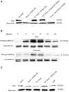
Incubation with recombinant Wnt5a, but not recombinant Wnt3a nor EGF enhanced villin protein expression. (A) In WT Ror2-transfected HT29 cells, villin protein was slightly increased compared to Wnt5a CM-treated mock-transfected cells. There was a noticeable increase in similarly treated WT Ror2-overexpressing cells. Incubation with rWnt5a (200 ng/mL) for 24 h enhanced villin expression to a similar degree. Treatment with rWnt3a (200 ng/mL) for 24 h did not induce any immunoreactive villin expression. (B) Challenge with EGF (100 nM) for 0, 2, 5, 10, 30, 60 min induced transient and robust increases in ERK1/2 phosphorylation in HT29 cells. In contrast, weak pERK1/2 immunoreactivity was detected in cells which had been challenged with low serum media, which did not contain EGF, over similar time points. (C) As previously observed, in HT29 cells which were mock-transfected or left untreated, low levels of villin protein expression were detected. In WT Ror2-overexpressing cells challenged with Wnt5a conditioned media for 24 h, villin protein expression was increased. In WT Ror2-overexpressing cells, challenged with EGF for a similar 24 h time period, low levels of villin expression were detectable, comparable to that found in mock-transfected or untreated control cells. β-actin immunoblotting was included as a loading control. These results are representative of three separately performed experiments.
Figure 7
Figure 7
Wnt5a challenge stimulated villin expression, via an ERK1/2-dependent mechanism in WT Ror2-overexpressing human intestinal epithelial cells (HIECs). (A) Villin protein was barely detectable in mock-transfected and/or unchallenged HIECs. Upon Wnt5a treatment, levels of villin transcript expression were slightly increased. Overexpression of WT Ror2 in these cells induced faint villin expression. Treating WT Ror2-cells with Wnt5a CM for 24 h noticeably enhanced villin protein expression. (B) Wnt5a addition in Ror2-overexpressing cells transiently and robustly increased dual-ERK1/2 phosphorylation over the course of an hour. (C) The Wnt5a-enhanced villin protein expression was prevented in similarly treated cells, in the presence of PD 098059 (10 μM). (D) However, increased villin protein levels were maintained in Wnt5a-challenged WT Ror2-cells, despite the presence of either Bisindolylmaleimide I (1 μM) or SB 203580 (10 μM). Yet, in the presence of D4476 (100 μM), Wnt5a-induced villin protein expression was prevented For all experiments described, Western blotting for immunoreactive β-actin was performed to demonstrate equal loading. These experiments reflect results from similar experiments performed separately.
Figure 8
Figure 8
Wnt5a treatment did not stimulate villin protein expression in 5YF Ror2-expressing HIECs but did induce villin protein expression in BDB Ror2-expressing HIECs. (A) In untreated WT Ror2-expressing cells, villin protein expression was not detectable; upon Wnt5a challenge for 24 h, villin expression was substantially increased. Levels of villin protein were low in untreated and Wnt5a-treated 5YF Ror2-expressing cells. Similar Wnt5a treatment upregulated the amount of immunoreactive villin in cells transfected with BDB Ror2. β-actin immunoreactivity was assessed to confirm equivalent loading. These results are representative of three separate experiments. (B) Incubation with recombinant Wnt3a protein did not stimulate villin protein expression in Ror2-overexpressing HIECs. Villin protein expression was not detectable in untreated mock-transfected HIEC, but was slightly enhanced in untreated WT Ror2- expressing cells. Villin protein expression was noticeably increased with Wnt5a CM-challenge for 24 h in WT Ror2-overexpressing cells. In similarly transfected cells, rWnt3a incubation for a 24-h period did not induce increases in villin protein. β-actin immunoreactivity was assessed to confirm equivalent loading. These results are representative of three separate experiments.
Figure 9
Figure 9
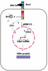
Model of Wnt-induced villin protein expression in adenocarcinoma-derived HT29 cells and fetally derived, untransformed human intestinal epithelial cells. Wnt5a interaction with Ror2 is mediated by the cysteine-rich domain. Phosphorylation of the tyrosines within in the Ser/Thr-1 region, possibly by Src, induces ERK1/2 phosphorylation. Stimulated ERK1/2 upregulates villin transcript expression; this subsequently increases villin protein. This Wnt5a-induced villin expression is also mediated by CK1. As well, CDX2, acting downstream of, or in parallel with, the activated ERK1/2 MAPK cascade could be mediating Wnt5a-induced increases in villin protein.

Similar articles

Cited by

References

    1. Abdullah H. I., Pedraza P. L., McGiff J. C., Ferreri N. R. (2008). CaR activation increases TNF production by mTAL cells via a Gi-dependent mechanism. Am. J. Physiol. Renal Physiol. 294, F345–F35410.1152/ajprenal.00509.2006 - DOI - PubMed
    1. Akbarzadeh S., Wheldon L. M., Sweet S. M., Talma S., Mardakheh F. K., Heath J. K. (2008). The deleted in brachydactyly B domain of ROR2 is required for receptor activation by recruitment of Src. PLoS ONE 3, e1873.10.1371/journal.pone.0001873 - DOI - PMC - PubMed
    1. Alessi D. R., Cuenda A., Ohen P., Udley D. T., Altiel A. R. (1995). PD 098059 is a specific inhibitor of the activation of mitogen-activated protein kinase kinase in vitro and in vivo. J. Biol. Chem. 270, 27489–2749410.1074/jbc.270.46.27489 - DOI - PubMed
    1. Aliaga J. C., Deschenes C., Beaulieu J. F., Calvo E. L., Rivard N. (1999). Requirement of the MAP kinase cascade for cell cycle progression and differentiation of human intestinal cells. Am. J. Physiol. 277, G631–G641 - PubMed
    1. Billiard J., Way D. S., Seestaller-Wehr L. M., Moran R. A., Mangine A., Bodine P. V. (2005). The orphan receptor tyrosine kinase Ror2 modulates canonical Wnt signaling in osteoblastic cells. Mol. Endocrinol. 19, 90–10110.1210/me.2004-0153 - DOI - PubMed