. 2011 Sep;7(9):e1002245.
doi: 10.1371/journal.ppat.1002245. Epub 2011 Sep 15.

A viral ubiquitin ligase has substrate preferential SUMO targeted ubiquitin ligase activity that counteracts intrinsic antiviral defence

Affiliations

A viral ubiquitin ligase has substrate preferential SUMO targeted ubiquitin ligase activity that counteracts intrinsic antiviral defence

Chris Boutell et al. PLoS Pathog. 2011 Sep.

Abstract

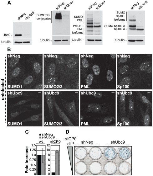
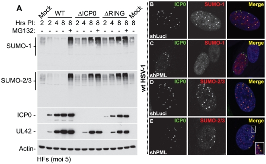
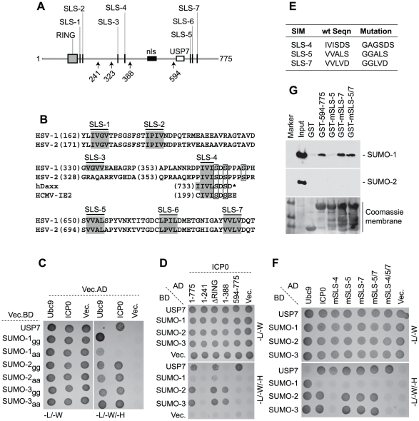
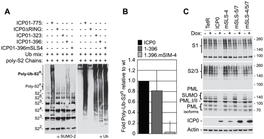
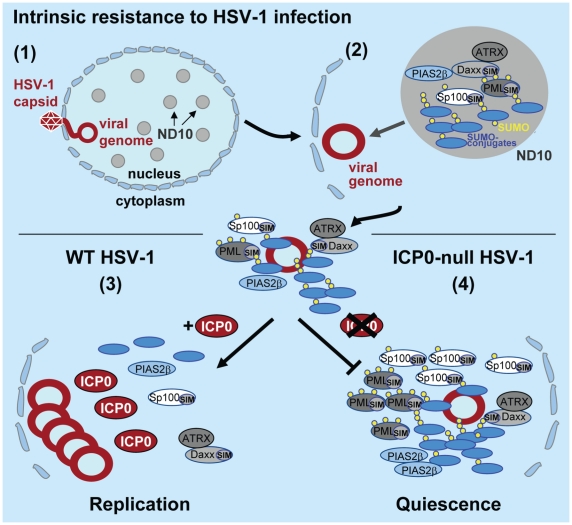
Intrinsic antiviral resistance represents the first line of intracellular defence against virus infection. During herpes simplex virus type-1 (HSV-1) infection this response can lead to the repression of viral gene expression but is counteracted by the viral ubiquitin ligase ICP0. Here we address the mechanisms by which ICP0 overcomes this antiviral response. We report that ICP0 induces the widespread proteasome-dependent degradation of SUMO-conjugated proteins during infection and has properties related to those of cellular SUMO-targeted ubiquitin ligases (STUbLs). Mutation of putative SUMO interaction motifs within ICP0 not only affects its ability to degrade SUMO conjugates, but also its capacity to stimulate HSV-1 lytic infection and reactivation from quiescence. We demonstrate that in the absence of this viral countermeasure the SUMO conjugation pathway plays an important role in mediating intrinsic antiviral resistance and the repression of HSV-1 infection. Using PML as a model substrate, we found that whilst ICP0 preferentially targets SUMO-modified isoforms of PML for degradation, it also induces the degradation of PML isoform I in a SUMO modification-independent manner. PML was degraded by ICP0 more rapidly than the bulk of SUMO-modified proteins in general, implying that the identity of a SUMO-modified protein, as well as the presence of SUMO modification, is involved in ICP0 targeting. We conclude that ICP0 has dual targeting mechanisms involving both SUMO- and substrate-dependent targeting specificities in order to counteract intrinsic antiviral resistance to HSV-1 infection.


Conflict of interest statement

The authors have declared that no competing interests exist.

Figures

Figure 1
Figure 1. The SUMO pathway contributes to intrinsic antiviral resistance to HSV-1 infection.
(A) Western blots analyzing the expression of Ubc9, SUMO-2/3 conjugates, PML and Sp100 in HFs stably expressing control (shNeg) or Ubc9 (shUbc9) shRNAs. (B) Nuclear distribution of endogenous SUMO-1 and SUMO-2/3 conjugates, PML and Sp100 at ND10 in uninfected shNeg and shUbc9 cells. Scale bar represents 5 µm. (C) shNeg or shUbc9 cells were infected with wt or ICP0-null mutant (ΔICP0) viruses expressing a β-galactosidase reporter gene from the tk locus. 24 hours post-infection the cells were stained for β-galactosidase expression and relative plaque formation efficiencies calculated and expressed as fold increase with respect to the appropriate control infection. Means and standard deviations of three independent experiments are presented. (D) Duplicate wells of shNeg and shUbc9 cells infected with two input doses of ΔICP0 virus and stained for β-galactosidase expression 24 hours post-infection.
Figure 2
Figure 2. ICP0 localizes to and induces the degradation of SUMO-1 and SUMO-2/3 conjugates during infection in a RING finger- and proteasome-dependent manner.
(A) HFs were infected at a MOI of 5 plaque forming units (pfu) per cell with wt HSV-1, ICP0-null (ΔICP0) or ICP0 RING finger deletion (ΔRING) mutant viruses in the absence or presence (-/+) of the proteasome inhibitor MG132. Cells were harvested at the indicated time points post-infection (Hrs PI) and analyzed for SUMO-1 and SUMO-2/3 abundance. The blots were reprobed for viral antigens ICP0 and UL42, and actin as a loading control. Longer exposures of western blots from duplicate experiments are shown in Figure S2B. (B–E) ICP0 localizes to SUMO-2/3, but not SUMO-1, conjugates in a PML-independent manner. Control (shLuci) or PML-depleted (shPML) HFs were infected with wt HSV-1 and the localization of ICP0 (green) to endogenous SUMO-1 and SUMO-2/3 conjugates (red) was analyzed in cells at the periphery of developing plaques (PML localization within these cells is shown in Figure S4). Nuclei were stained with DAPI. The insert at the lower right corner shows an expanded region highlighted by the white box.
Figure 3
Figure 3. ICP0 interacts with SUMO in a SIM-dependent manner.
(A) Locations of predicted SIM-Like Sequences (SLS, black vertical bars), the RING finger (grey box), nuclear localization sequence (nls, black horizontal bar), and USP7 binding domain (white box) within ICP0. Number arrows refer to amino acid coordinates. (B) Alignment of SLS in HSV-1 and HSV-2 ICP0. Grey boxes highlight the hydrophobic core within individual SLSs. SLS-4 is additionally aligned with SIMs of hDaxx and HCMV IE2. Square boxes adjacent to SLS-4 highlight phosphorylated serine residues. (C) Y2H analysis showing ICP0 interaction with SUMO-2/3, but not SUMO-1, in a C-terminal di-glycine independent manner. Ubc9 and USP7 are positive controls for SUMO and ICP0 interaction respectively. Mated diploids were plated out onto media lacking leucine and tryptophan (-L/-W, indicating the presence of both plasmids) or leucine, tryptophan, and histidine (-L/-W/-H). Positive interactions are indicated by growth on medium lacking histidine. GAL4 activation domain (AD) or binding domain (BD) fusion orientations are highlighted. Vec indicates the respective empty vector control. (D) ICP0 interacts with SUMO-2/3 in a RING finger-independent manner and requires residues between amino acids 241 to 388. (E) Table highlighting residue mutations made within SLS-4, -5 and -7. (F) Mutation of SLS-4 inhibits ICP0’s ability to interact with SUMO-2/3 in Y2H assay. (G) GST pull down assay demonstrating that the C-terminal third of ICP0 (residues 594-775) interacts with SUMO-1.
Figure 4
Figure 4. SLS-4 is required for the in vitro ubiquitination of poly-SUMO-2 chains and degradation of SUMO conjugates in vivo.
(A) In vitro ubiquitination reactions were carried out in the presence of E1, UbcH5a and ubiquitin (Ubmix), poly-SUMO-2 chains and purified wt ICP0, ICP0ΔRING, or ICP0 C-terminal truncation mutants ICP01-323, ICP01-396 or ICP01-396mSLS-4 as indicated (-/+). Ubiquitinated products were analyzed by western blot for ubiquitinated SUMO species (left-) and poly-ubiquitin chain formation (right-hand panels, respectively). Poly-ubiquitinated SUMO-2 species are labelled Poly-Ub-S2n. Superscripts denote the number of SUMO-2 (S2) molecules within the chain. (B) Quantification of Poly-Ub-S2n species (dotted line) detected by western blot analysis in reaction mixtures containing ICP0, ICP01-396, and ICP01-396mSLS-4 expressed as a relative fold decrease in relation to reaction mixtures containing wt ICP0. Error bars represent the standard deviation in Poly-Ub-S2n levels detected over four independent experiments. (C) Analysis of SUMO conjugates and PML stability in cells induced to express wt ICP0, ICP0 mSLS-4, -5/7, -4/5/7 mutants, or empty vector control (TetR) cells at 24 hours post-induction with doxycycline (Dox; +) compared to uninduced controls (-). The actin and ICP0 blots provide loading and ICP0 expression controls.
Figure 5
Figure 5. ICP0 preferentially induces the degradation of SUMO-conjugated PML.
(A) Maps of PML.I and PML.IV depicting RING, B-box, coiled-coil and nls motifs. Arrows indicate the SIM and lysine (K) residues mutated in this study, vertical lines show exon boundaries in PML.I, and the black horizontal bar represents the PML.IV specific exon 8b. (B) ICP0 preferentially induces the proteasome-dependent degradation of SUMO-modified forms of PML.I-VI. Cells expressing EYFP-PML isoforms I-VI were infected with wt (MOI 2) or ICP0-null mutant HSV-1 (ΔICP0, MOI 10), in the presence or absence of MG132, and harvested at 6 hours post-infection. Cell extracts were analyzed by western blot for EYFP-PML in comparison with mock-infected controls. (C and D) ICP0 degrades EYFP-PML.I but not EYFP-PML.IV K160/490R mutants in both control cells (C) and cells depleted of endogenous PML (D). (E) ICP0 induces the degradation of EYFP-PML.I.K65/160/490/616R in a SUMO modification-independent manner.
Figure 6
Figure 6. SLSs within ICP0 regulate its ability to complement and reactivate mutant HSV-1 viruses in cell culture.
(A) As in Figure 3A. (B) Complementation of ICP0-null mutant HSV-1 plaque formation by prior induction of expression of various N-terminal fragments of ICP0 (as depicted in A) and full-length ICP0 carrying individual or combined SLS mutations in the inducible cell line system. The titre of a mutant virus stock was determined in each cell line and plotted with respect to that in cells expressing wt ICP0. Means and standard deviations of two to seven independent determinations are presented. (C) Analysis of ICP0 induced reactivation of gene expression from quiescent HSV-1 genomes. Cells were infected with multiply defective HSV-1 mutant in1374 to establish quiescently infected cultures, then 24 h later ICP0 expression was induced with doxycycline. Reactivation was assessed the following day by staining for β-galactosidase expression from the marker gene in the in1374 genome. The proportion of reactivated cells in each cell line was expressed as a percentage of that in cells expressing wt ICP0 following determination of positive cell numbers in three high magnification views of each sample. Means and standard deviations are presented.
Figure 7
Figure 7. ICP0 and its viral orthologues induce the degradation of SUMO conjugates independently of virus infection and the degradation of Ubc9.
(A) Schematic representation of ICP0 orthologue proteins BICP0, EICP0, VICP0 and PICP0 illustrating the distribution of putative SIM-Like Sequence (SLS) motifs in relation to their respective RING finger domains (grey boxes). Numbering reflects the first and last amino acid within each ORF. (B) Amino acid sequence of putative SLS motifs identified within each ICP0 viral orthologue. Numbers refer to the coordinates of the first amino acid shown within each SLS with respect to their individual ORF sequence. Bold lettering represents motifs that conform to the SIM consensus as described –. Boxed sequences show conserved triplets composed of serine or acidic residues. (C) Cell lines induced to express ICP0, myc-tagged viral orthologues (BICP0, EICP0 and PICP0), or control cells (TetR) were analyzed for Ubc9 expression levels in relation to SUMO conjugate abundance 24 hours after treatment with doxycycline (0.1 µg/ml) (−/+). Blots were reprobed for ICP0, myc-tagged orthologue expression, and tubulin as a loading control.
Figure 8
Figure 8. Model depicting the regulation of intrinsic antiviral resistance to HSV-1 infection mediated by the SUMO conjugation pathway.
(1) During the initial stages of HSV-1 infection viral genomes enter the nucleus of infected cells. (2) Major ND10 components including PML, Sp100, hDaxx, and ATRX are recruited into foci that are closely associated with incoming HSV-1 genomes. This recruitment is dependent upon the SUMO conjugation pathway (Figures 1 and S1) and SIMs within PML, Sp100 and hDaxx . (3) During wt HSV-1 infection, the STUbL-like activity of ICP0 promotes the preferential degradation of SUMO-conjugated proteins leading to the dispersal of restriction factors and the efficient onset of viral replication. (4) In the absence of ICP0, specific SUMO-conjugated proteins mediate the transcriptional repression of viral gene expression leading to the establishment of viral quiescence and latency.

References

    1. Randow F, Lehner PJ. Viral avoidance and exploitation of the ubiquitin system. Nat Cell Biol. 2009;11:527–534. - PubMed
    1. Everett RD. The roles of ICP0 during HSV-1 infection. Alpha herpesviruses: molecular and cellular biology. In: Sandri-Goldin RM, editor. Wymondham, United Kingdom: Caister Academic Press; 2006b. pp. 39–64.
    1. Boutell C, Sadis S, Everett RD. Herpes simplex virus type 1 immediate-early protein ICP0 and is isolated RING finger domain act as ubiquitin E3 ligases in vitro. J Virol. 2002;76:841–850. - PMC - PubMed
    1. Lium EK, Silverstein S. Mutational analysis of the herpes simplex virus type 1 ICP0 C3HC4 zinc ring finger reveals a requirement for ICP0 in the expression of the essential alpha27 gene. J Virol. 1997;71:8602–8614. - PMC - PubMed
    1. Everett RD. A detailed mutational analysis of Vmw110, a trans-acting transcriptional activator encoded by herpes simplex virus type 1. Embo J. 1987;6:2069–2076. - PMC - PubMed

Publication types

MeSH terms