. 2011 Sep;7(9):e1002285.
doi: 10.1371/journal.pgen.1002285. Epub 2011 Sep 15.

Inactivation of Pmel alters melanosome shape but has only a subtle effect on visible pigmentation

Affiliations

Inactivation of Pmel alters melanosome shape but has only a subtle effect on visible pigmentation

Anders R Hellström et al. PLoS Genet. 2011 Sep.

Abstract

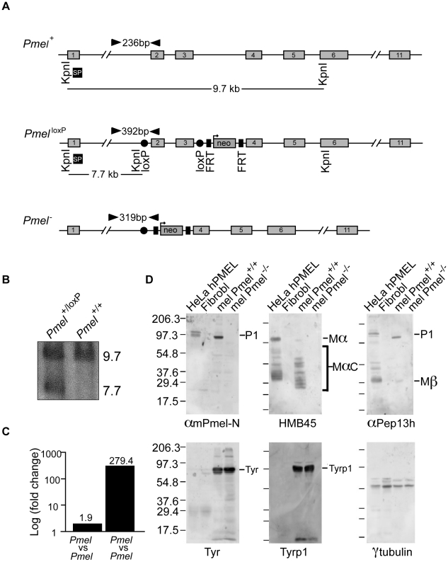
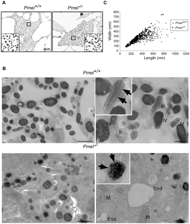
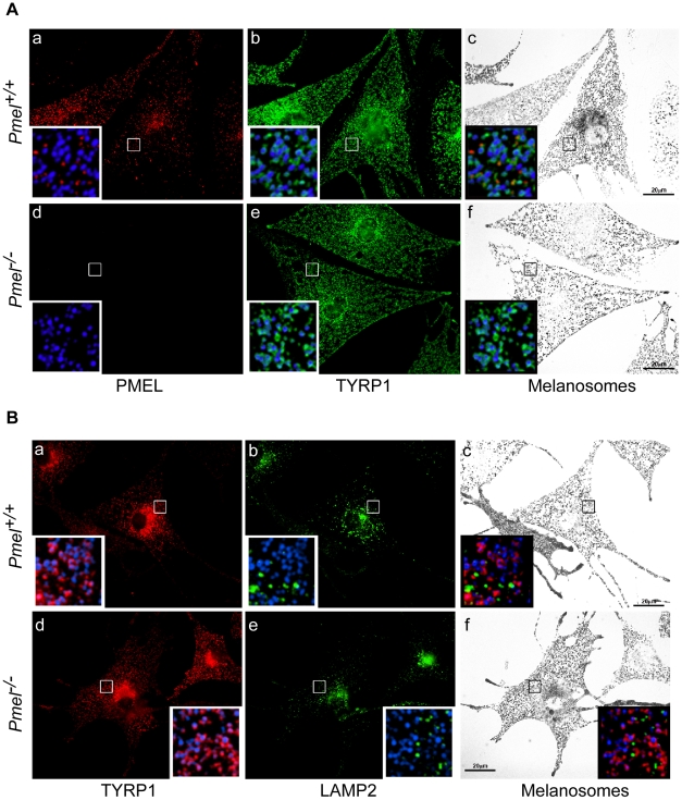
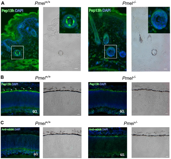
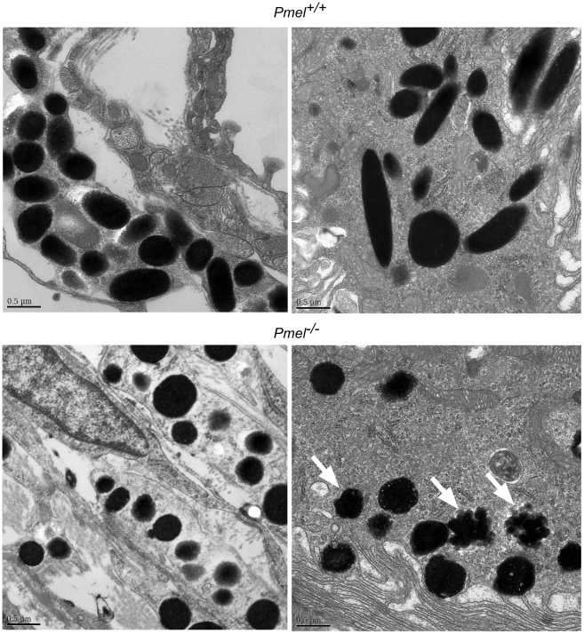
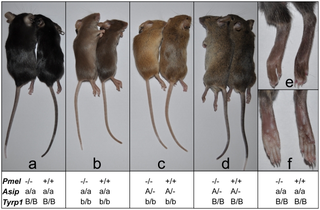
PMEL is an amyloidogenic protein that appears to be exclusively expressed in pigment cells and forms intralumenal fibrils within early stage melanosomes upon which eumelanins deposit in later stages. PMEL is well conserved among vertebrates, and allelic variants in several species are associated with reduced levels of eumelanin in epidermal tissues. However, in most of these cases it is not clear whether the allelic variants reflect gain-of-function or loss-of-function, and no complete PMEL loss-of-function has been reported in a mammal. Here, we have created a mouse line in which the Pmel gene has been inactivated (Pmel⁻/⁻). These mice are fully viable, fertile, and display no obvious developmental defects. Melanosomes within Pmel⁻/⁻ melanocytes are spherical in contrast to the oblong shape present in wild-type animals. This feature was documented in primary cultures of skin-derived melanocytes as well as in retinal pigment epithelium cells and in uveal melanocytes. Inactivation of Pmel has only a mild effect on the coat color phenotype in four different genetic backgrounds, with the clearest effect in mice also carrying the brown/Tyrp1 mutation. This phenotype, which is similar to that observed with the spontaneous silver mutation in mice, strongly suggests that other previously described alleles in vertebrates with more striking effects on pigmentation are dominant-negative mutations. Despite a mild effect on visible pigmentation, inactivation of Pmel led to a substantial reduction in eumelanin content in hair, which demonstrates that PMEL has a critical role for maintaining efficient epidermal pigmentation.


Conflict of interest statement

The authors have declared that no competing interests exist.

Figures

Figure 1
Figure 1. Strategy for the generation and validation of Pmel−/− mice.
(A) Schematic representation of the Pmel+, PmelloxP, and Pmel alleles. The exons (gray boxes), primers used for genotyping (black arrowheads), KpnI restriction cleavage sites and the target site of Southern blot probe (SP, black box) are all indicated. In the PmelloxP allele, exon 2 and exon 3 are loxP-flanked (red circles) and a neomycin (green box) gene is flanked by two FRT-sites (blue boxes). In the Pmel allele, exon 2 and exon 3 are deleted after Cre-recombinase induced homologous recombination between the loxP sites. (B) Southern blot showing targeted and control ES-cells. The wild-type and the PmelloxP alleles generate a 9.7 kb band and a 7.7 kb band, respectively. (C) Pmel mRNA expression. A 1.9-fold change was detected in skin tissue when homozygous Pmel+/+ mice were compared to Pmel +/− heterozygotes. A 279-fold change was detected when homozygous Pmel −/− mice were compared to homozygous wild-type mice. (D) Western blot analysis. Whole cell lysates from primary cultures of skin-derived melanocytes from wild-type (mel Pmel+/+) or Pmel −/− (mel Pmel−/−) C57BL/6 mice were used, and lysates from transfected HeLa cells expressing human PMEL (HeLa hPMEL) or untransfected human fibroblasts (Fibrobl) were used as positive and negative controls. The lysates were fractionated by SDS-PAGE and analyzed by immunoblotting with a panel of antibodies directed against PMEL (αmPmel-N, HMB45, and αPep13h) or the control proteins, tyrosinase (TYR), tyrosinase-related protein 1 (TYRP1), and γ-tubulin. αmPmel-N recognizes an N-terminal peptide from mouse PMEL; HMB45 recognizes an epitope derived from the central region of PMEL that is enriched on fibrils; and αPep13h detects the PMEL C-terminus. Migration of molecular weight markers (in kDa) is shown to the left, and relevant bands are indicated to the right.
Figure 2
Figure 2. Morphology of melanosomes observed in primary cultures of skin-derived melanocytes from wild-type C57BL/6 (Pmel+/+) and Pmel−/−mice.
(A) Bright-field microscopy; scale bar, 20µm; inset, 5X magnification. Note that the distribution of melanosomes is similar in both sets of melanocytes. (B) Electron microscopy. Cross-sections of melanosomes in the Pmel+/+ melanocytes are both spherical and rod shaped, whilst they are only spherical in the Pmel −/− melanocytes. I, II, III, and IV represent stage I, II, III, and IV melanosomes. M, mitochondria; End, endosomes. Note the presence of fibrillar stage II and III melanosomes in the wild-type melanocytes (panel Bb, marked with arrowheads in inset) in contrast to the granular deposits of melanin in Pmel −/− melanosomes (panel Bd, marked with arrows in inset). Bars: 500 nm and 200 nm, in left and right panels respectively (C) Plot of the diameters of melanosomes along the long (length) and short (width) axes measured from electron microscopy images; n>200 for both genotypes. There was a weak correlation between the length and width of melanosomes in Pmel+/+ cells (r = 0.52), consistent with their ellipsoidal shape, whereas in Pmel −/− cells, there was a strong correlation (r = 0.93), indicating a spherical shape. The difference in the length/width ratio between wild-type and Pmel −/− is overwhelmingly significant (Analysis of variance; F = 161, d.f.1 = 1, d.f.2 = 451; P<10−6).
Figure 3
Figure 3. Immunofluorescence and bright field microscopy of primary melanocytes from C57BL/6 wild-type (Pmel+/+) and knockout (Pmel −/−) mice.
(A) Cells immunolabeled with antibodies against the mature PMEL protein (HMB45, red; a, d, insets) and TYRP1 (green; b, e, insets); the melanosomes were visualized by bright field (c, f) and pseudo-colored blue in the insets. Note the presence of TYRP1 surrounding melanosomes in both sets of melanocytes. (B) Cells immunolabeled with antibodies against the late endosome/ lysosome marker LAMP2 (green; b, e, insets) and TYRP1 (red; a, d, insets); the melanosomes were visualized by bright field (c, f) and pseudo-colored blue in the insets. Note the segregation of LAMP2 labeling from pigment granules labeled by TYRP1. Size bar, 20 µm.
Figure 4
Figure 4. Immunohistochemistry of skin and retina from Pmel+/+and Pmel−/−mice.
Sections of skin (A) or retina (B, C) from Pmel+/+ (a, b) and Pmel −/− (c, d) mice were immunolabeled with αPep13h (anti-PMEL) (A, B) or secondary antibody alone (C) and counterstained with DAPI to label nuclei, and analyzed by immunofluorescence microscopy (a, c) or bright field microscopy to assess pigmentation (b, d). (A) PMEL-immunoreactive cells were found in the hair follicles of the skin in wild-type mice (a–b), but not in the Pmel −/− mice (c–d). Insets, two-fold enlargement of the boxed region. (B) αPep13h immunoreactive cells (green) were found in the choroid (arrowheads) and in the RPE from the Pmel+/+ mouse (a), but only in the RPE/ Bruch's membrane from the Pmel −/− mouse (c). GCL, ganglion cell layer. (C) Immunostaining obtained using only the secondary anti-rabbit antibody shows a signal from Bruch's membrane. Size bar, 20 µm.
Figure 5
Figure 5. Electron microscopy of choroidal (upper and lower left micrographs) and RPE cells (upper and lower right micrographs), including melanosomes, from wild-type C57BL/6 mice (upper two micrographs) and Pmel−/− mice (lower two micrographs), respectively.
A scale bar is depicted in the lower left hand corner of each micrograph. Melanosomes of both choroidal and RPE cells of the affected animal appear spherical with irregular borders, the latter more obvious in the RPE cells (lower right micrographs, indicated by white arrowheads). Further, no cigar-shaped melanosomes were found in either choroidal or RPE cells in the Pmel −/− mice.
Figure 6
Figure 6. Phenotypic effects on pigmentation of the Pmel−/− genotype on four different genetic backgrounds.
Coat color was assessed in different genetic backgrounds by pairwise comparisons from an F2 intercross generation. Within each pair, the Pmel −/− and Pmel+/+ mice are to the left and right, respectively. (a) The non-agouti black (Asipa/a) Pmel −/− mice displayed a subtle dilution of coat color and tail skin. (b) PMEL inactivation in brown mice (Asipa/a Tyrp1b/b) caused the most dramatic reduction in coat color and tail skin pigmentation. (c) The black hairs of the Pmel −/− mice on brown agouti (AsipA/− Tyrp1b/b) background was diluted giving the coat a more yellow/light brown appearance compared to the wild-type. (d) The weakest phenotypic effect associated with the Pmel −/− genotype was seen in agouti mice (AsipA/−) but the coat and skin color were slightly lighter relative to the wild-type. (e, f) The skin of the feet of non-agouti black (Asipa/a) Pmel −/− mice were less pigmented compared to the wild-type.

Similar articles

Cited by

References

    1. Ito S, Wakamatsu K. Chemistry of mixed melanogenesis--pivotal roles of dopaquinone. Photochem Photobiol. 2008;84:582–592. - PubMed
    1. Furumura M, Sakai C, Potterf SB, Vieira WD, Barsh GS, et al. Characterization of genes modulated during pheomelanogenesis using differential display. Proc Natl Acad Sci U S A. 1998;95:7374–7378. - PMC - PubMed
    1. Baxter LL, Pavan WJ. Pmel17 expression is Mitf-dependent and reveals cranial melanoblast migration during murine development. Gene Expr Patterns. 2003;3:703–707. - PubMed
    1. Theos AC, Truschel ST, Raposo G, Marks MS. The Silver locus product Pmel17/gp100/Silv/ME20: controversial in name and in function. Pigment Cell Res. 2005;18:322–336. - PMC - PubMed
    1. Chakraborty AK, Platt JT, Kim KK, Kwon BS, Bennett DC, et al. Polymerization of 5,6-dihydroxyindole-2-carboxylic acid to melanin by the pmel 17/silver locus protein. Eur J Biochem. 1996;236:180–188. - PubMed

Publication types

MeSH terms