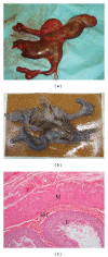
Bipartite anterior extraperitoneal teratoma: evidence for the embryological origins of teratomas?
- PMID: 21949666
- PMCID: PMC3163403
- DOI: 10.1155/2011/208940
Bipartite anterior extraperitoneal teratoma: evidence for the embryological origins of teratomas?
Abstract
Teratomas are thought to arise from totipotent primordial germ cells (PGCs) Dehner (1983) which may miss their target destination Moore and Persaud (1984). Teratomas can occur anywhere from the brain to the coccygeal area but are usually in the midline close to the embryological position of the gonadal ridges Bale (1984), Nguyen and Laberge (2000). We report a case of a bipartite anterior extraperitoneal teratoma. This is an unusual position for a teratoma, but one which may support the "missed target" theory of embryology.
Figures


Similar articles
-
Neonatal teratomas.Early Hum Dev. 2010 Oct;86(10):643-7. doi: 10.1016/j.earlhumdev.2010.08.016. Epub 2010 Sep 29. Early Hum Dev. 2010. PMID: 20884137 Review.
-
Ovarian and non-ovarian teratomas: a wide spectrum of features.Jpn J Radiol. 2021 Feb;39(2):143-158. doi: 10.1007/s11604-020-01035-y. Epub 2020 Sep 1. Jpn J Radiol. 2021. PMID: 32875471 Review.
-
Teratoma involving adrenal gland - A case report and review of literature.Indian J Radiol Imaging. 2019 Oct-Dec;29(4):472-476. doi: 10.4103/ijri.IJRI_452_18. Epub 2019 Dec 31. Indian J Radiol Imaging. 2019. PMID: 31949356 Free PMC article.
-
Lower lip teratoma with ventral capillary malformation in an infant: case report and literature review.Int J Oral Maxillofac Surg. 2009 Dec;38(12):1330-3. doi: 10.1016/j.ijom.2009.06.018. Epub 2009 Jul 23. Int J Oral Maxillofac Surg. 2009. PMID: 19631510 Review.
-
Germ cell tumors of the gonads: a selective review emphasizing problems in differential diagnosis, newly appreciated, and controversial issues.Mod Pathol. 2005 Feb;18 Suppl 2:S61-79. doi: 10.1038/modpathol.3800310. Mod Pathol. 2005. PMID: 15761467 Review.
Cited by
-
Types and Frequency of Neural Elements in Mature Ovarian Teratomas: An 11-Year Study from Rural India.J Microsc Ultrastruct. 2022 Dec 1;10(4):154-158. doi: 10.4103/jmau.jmau_94_20. eCollection 2022 Oct-Dec. J Microsc Ultrastruct. 2022. PMID: 36687327 Free PMC article.
References
-
- Dehner LP. Gonadal and extragonadal germ cell neoplasia of childhood. Human Pathology. 1983;14(6):493–511. - PubMed
-
- Moore K, Persaud T. The Developing Human. Philadelphia, Pa, USA: WB Saunders; 1993.
-
- Bale PM. Sacrococcygeal developmental abnormalities and tumors in children. Perspectives in Pediatric Pathology. 1984;8(1):9–56. - PubMed
-
- Nguyen LT, Laberge J-M. Teratomas, dermoids and other soft tissue tumors. In: Ashcraft KW, editor. Pediatric Surgery. Philadelphia, Pa, USA: Saunders; 2000. pp. 972–974.
-
- de Lagausie P, de Napoli SC, Stempfle N, et al. Highly differentiated teratoma and fetus-in-fetu: a single pathology? Journal of Pediatric Surgery. 1997;32(1):115–116. - PubMed
Publication types
LinkOut - more resources
Full Text Sources

