. 2011;6(9):e23309.
doi: 10.1371/journal.pone.0023309. Epub 2011 Sep 16.

Erythrocyte and porcine intestinal glycosphingolipids recognized by F4 fimbriae of enterotoxigenic Escherichia coli

Affiliations

Erythrocyte and porcine intestinal glycosphingolipids recognized by F4 fimbriae of enterotoxigenic Escherichia coli

Annelies Coddens et al. PLoS One. 2011.

Abstract

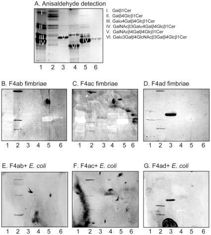
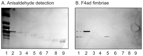
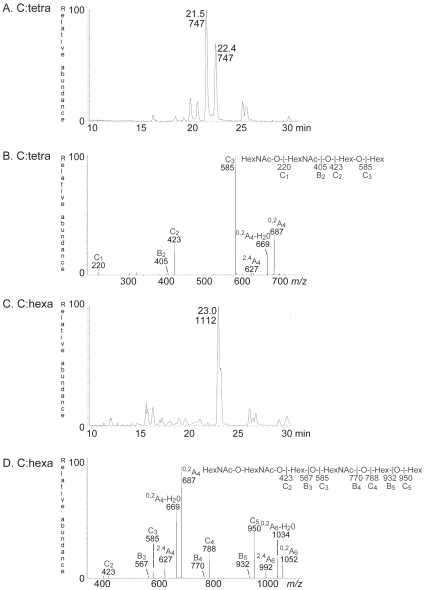
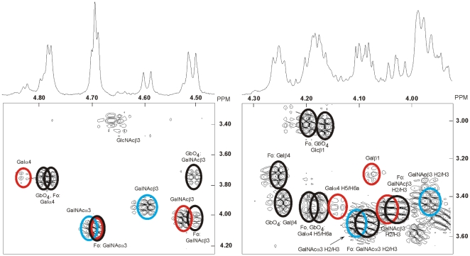
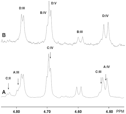
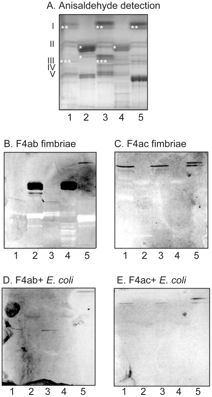
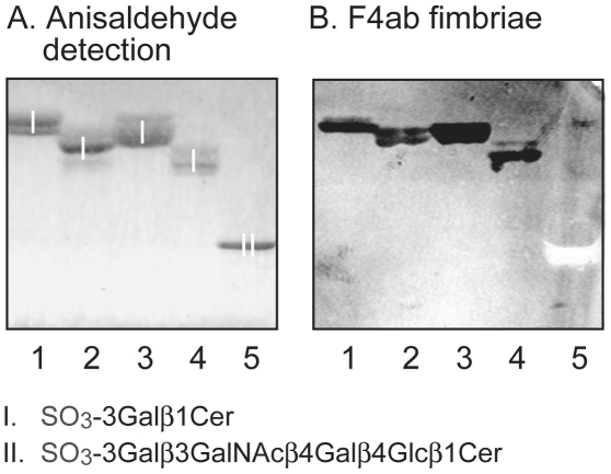
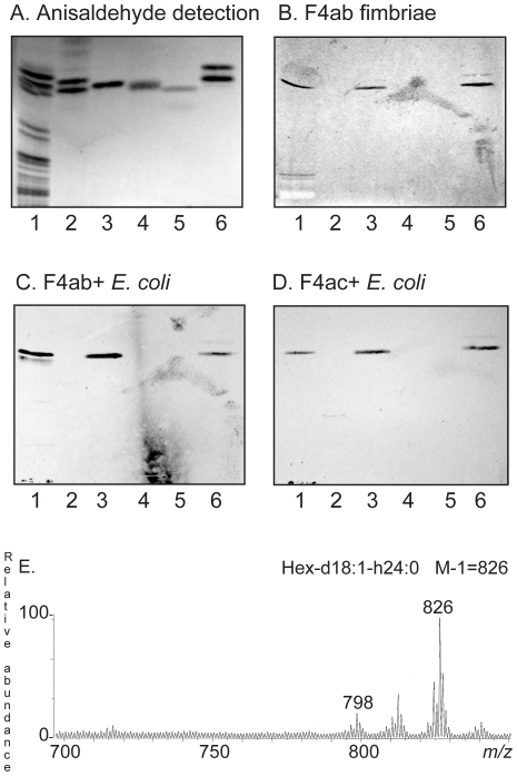
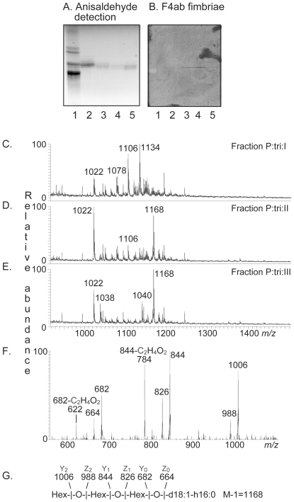
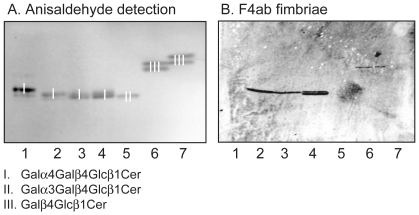
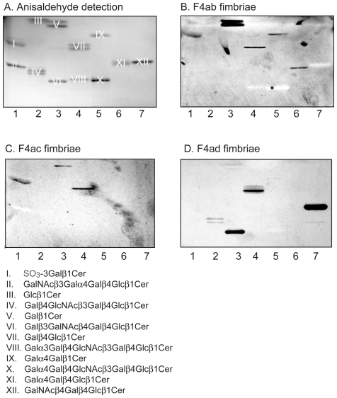
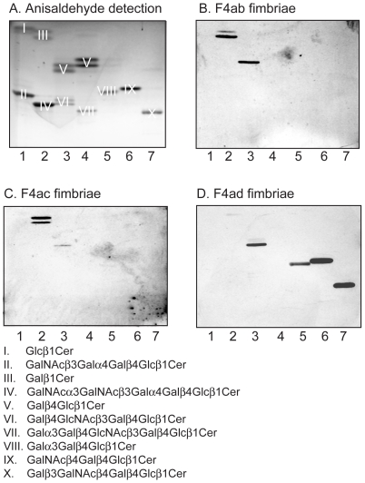
Enterotoxigenic F4-fimbriated Escherichia coli is associated with diarrheal disease in neonatal and postweaning pigs. The F4 fimbriae mediate attachment of the bacteria to the pig intestinal epithelium, enabling an efficient delivery of diarrhea-inducing enterotoxins to the target epithelial cells. There are three variants of F4 fimbriae designated F4ab, F4ac and F4ad, respectively, having different antigenic and adhesive properties. In the present study, the binding of isolated F4ab, F4ac and F4ad fimbriae, and F4ab/ac/ad-fimbriated E. coli, to glycosphingolipids from erythrocytes and from porcine small intestinal epithelium was examined, in order to get a comprehensive view of the F4-binding glycosphingolipids involved in F4-mediated hemagglutination and adhesion to the epithelial cells of porcine intestine. Specific interactions between the F4ab, F4ac and F4ad fimbriae and both acid and non-acid glycosphingolipids were obtained, and after isolation of binding-active glycosphingolipids and characterization by mass spectrometry and proton NMR, distinct carbohydrate binding patterns were defined for each fimbrial subtype. Two novel glycosphingolipids were isolated from chicken erythrocytes, and characterized as GalNAcα3GalNAcß3Galß4Glcß1Cer and GalNAcα3GalNAcß3Galß4GlcNAcß3Galß4Glcß1Cer. These two compounds, and lactosylceramide (Galß4Glcß1Cer) with phytosphingosine and hydroxy fatty acid, were recognized by all three variants of F4 fimbriae. No binding of the F4ad fimbriae or F4ad-fimbriated E. coli to the porcine intestinal glycosphingolipids occurred. However, for F4ab and F4ac two distinct binding patterns were observed. The F4ac fimbriae and the F4ac-expressing E. coli selectively bound to galactosylceramide (Galß1Cer) with sphingosine and hydroxy 24:0 fatty acid, while the porcine intestinal glycosphingolipids recognized by F4ab fimbriae and the F4ab-fimbriated bacteria were characterized as galactosylceramide, sulfatide (SO(3)-3Galß1Cer), sulf-lactosylceramide (SO(3)-3Galß4Glcß1Cer), and globotriaosylceramide (Galα4Galß4Glcß1Cer) with phytosphingosine and hydroxy 24:0 fatty acid. Finally, the F4ad fimbriae and the F4ad-fimbriated E. coli, but not the F4ab or F4ac subtypes, bound to reference gangliotriaosylceramide (GalNAcß4Galß4Glcß1Cer), gangliotetraosylceramide (Galß3GalNAcß4Galß4Glcß1Cer), isoglobotriaosylceramide (Galα3Galß4Glcß1Cer), and neolactotetraosylceramide (Galß4GlcNAcß3Galß4Glcß1Cer).


Conflict of interest statement

Competing Interests: The authors have declared that no competing interests exist.

Figures

Figure 1
Figure 1. Binding of F4 fimbriae and F4-fimbriated Escherichia coli to erythrocyte non-acid glycosphingolipid mixtures.
Chemical detection by anisaldehyde (A), and autoradiograms obtained by binding of 125I-labeled F4ab fimbriae (B), F4ac fimbriae (C), F4ad fimbriae (D), and 35S-labeled F4ab-expressing E. coli (E), F4ac-expressing E. coli (F), and F4ad-expressing E. coli (G). The lanes were: Lane 1, non-acid glycosphingolipids of human erythrocytes blood group AB, 80 µg; Lane 2, non-acid glycosphingolipids of chicken erythrocytes, 40 µg; Lane 3, non-acid glycosphingolipids of guinea pig erythrocytes, 40 µg; Lane 4, non-acid glycosphingolipids of rabbit erythrocytes, 40 µg; Lane 5, non-acid glycosphingolipids of porcine erythrocytes, 40 µg; Lane 6, reference globotetraosylceramide (GalNAcß3Galα4Galß4Glcß1Cer) of human erythrocytes, 4 µg. The major compounds visualized with anisaldehyde in (A) are marked with Roman numbers, and the corresponding glycosphingolipid structures are given to the right of the chromatogram.
Figure 2
Figure 2. Binding of F4ad fimbriae to slow-migrating non-acid glycosphingolipid fractions isolated from chicken erythrocytes.
Chemical detection by anisaldehyde (A), and autoradiograms obtained by binding of 125I-labeled F4ad fimbriae (B). The lanes were: Lane 1, non-acid glycosphingolipids of chicken erythrocytes, 40 µg; Lanes 2–9, glycosphingolipid fractions isolated from chicken erythrocytes, 0.5–2 µg/lane.
Figure 3
Figure 3. Characterization of the F4 fimbriae binding tetra- and hexaglycosylceramide of chicken erythrocytes.
(A) Base peak chromatogram from LC-ESI/MS of the saccharide obtained by digestion with Rhodococcus endoglycoceramidase II of the F4-binding glycosphingolipid fraction C:tetra-II from chicken erythrocytes. (B) MS2 spectrum of the ion at m/z 747 (retention time 21.4 min). (C) Base peak chromatogram from LC-ESI/MS of the saccharide obtained by digestion with Rhodococcus endoglycoceramidase II of the F4-binding glycosphingolipid fraction C:hexa from chicken erythrocytes. (D) MS2 spectrum of the ion at m/z 1112 (retention time 23.0 min).
Figure 4
Figure 4. Proton NMR of the F4-binding glycosphingolipid (fraction C:tetra-I) from chicken erythrocytes.
Anomeric regions of the 600 MHz proton NMR of the F4-binding glycosphingolipid (fraction C:tetra-I) from chicken erythrocytes (30°C). The sample was dissolved in dimethyl sulfoxide-D2O (98∶2, by volume) after deuterium exchange. Below each section the corresponding DQF-COSY spectrum showing mainly the H1/H2 connectivities are displayed. Connectivities stemming from the same structure are color-coded by superimposed ellipses. Thus, the Forssman pentaglycosylceramide (Fo; GalNAcα3GalNAcß3Galα4Galß4Glcß1Cer) and globoside (GbO4; GalNAcß3Galα4Galß4Glcß1Cer) are black, whereas the two novel four-sugar compounds B and C are colored blue and red, respectively. Furthermore, only connectivities other than H1/H2 ones are denoted as such.
Figure 5
Figure 5. Proton NMR of fraction C:tetra-I and fraction C-tetra-II from chicken erythrocytes.
Low-field part of the anomeric regions of the 600 MHz spectra of fraction C:tetra-I (A) and fraction C-tetra-II (B). The arrows in (A) indicate which resonances that have disappeared in (B) and these are labeled according to the scheme in Table 1. Likewise, the remaining resonances in (B) are also labeled as in Table 1.
Figure 6
Figure 6. Binding of F4ab and F4ac fimbriae, and F4ab- and F4ac-fimbriated Escherichia coli, to mixtures of glycosphingolipids from porcine intestinal mucosa.
Chemical detection by anisaldehyde (A), and autoradiograms obtained by binding of F4ab fimbriae (B), F4ac fimbriae (C), F4ab-expressing E. coli (D), and F4ac-expressing E. coli (E). The lanes were: Lane 1, non-acid glycosphingolipids of 3-day old piglet small intestinal mucosa, 40 µg; Lane 2, acid glycosphingolipids of 3-day old piglet small intestinal mucosa, 20 µg; Lane 3, non-acid glycosphingolipids of adult pig 1 small intestinal mucosa, 40 µg; Lane 4, acid glycosphingolipids of adult pig 1 small intestinal mucosa, 40 µg; Lane 5, non-acid glycosphingolipids of adult pig 2 small intestinal mucosa, 40 µg. The Roman numbers to the left of panel A indicate the approximate number of carbohydrate residues in the bands in the non-acid fractions (lanes 1, 3 and 5). The approximate migration level of the F4ab-/F4ac-binding compounds have been marked with *, **, and *** in panel A.
Figure 7
Figure 7. Binding of F4ab fimbriae to reference glycosphingolipids.
Chemical detection by anisaldehyde (A), and autoradiograms obtained by binding of 125I-labeled F4ab fimbriae (B). The lanes were: Lane 1, sulfatide (SO3-3Galß1Cer) with d18:1-24:1 ceramide, 4 µg; Lane 2, sulfatide with d18:1-h16:0 ceramide, 4 µg; Lane 3, sulfatide with t18:0-h24:0 ceramide, 4 µg; Lane 4, sulf-lactosylceramide (SO3-3Galß4Glcß1Cer) with t18:0-h16:0 ceramide, 4 µg; Lane 5, reference sulf-gangliotetraosylceramide (SO3-3Galß3GalNAcß4Galß4Glcß1Cer), 4 µg. The glycosphingolipids visualized with anisaldehyde in (A) are marked with Roman numbers, and the corresponding glycosphingolipid structures are given below the chromatograms.
Figure 8
Figure 8. Characterization of the F4ab and F4ac binding monoglycosylceramides from porcine small intestinal mucosa.
Chemical detection by anisaldehyde (A), and autoradiograms obtained by binding of 125I-labeled F4ab fimbriae (B), 35S-labeled F4ab-expressing E. coli (C), and F4ac-expressing E. coli (D). The glycosphingolipids were separated using chloroform/methanol/water 65∶25∶4 (by volume) as solvent system. The lanes were: Lane 1, non-acid glycosphingolipids of adult pig 1 small intestinal mucosa, 40 µg; Lanes 2–5, monoglycosylceramides isolated from adult pig small intestinal mucosa, 4 µg/lane; Lane 6, reference galactosylceramide (Galß1Cer) with d18:1-h18:0-h24:0 ceramide, 4 µg. (E) Negative ion FAB mass spectrum of fraction P:mono from adult pig small intestinal mucosa. Above the spectrum is an interpretation formula representing the species with d18:1-h24:0 ceramide. The analysis was done as described in the “Materials and Methods” section.
Figure 9
Figure 9. Characterization of the F4ab fimbriae binding triglycosylceramides from porcine small intestinal mucosa.
Chemical detection by anisaldehyde (A), and autoradiogram obtained by binding of 125I-labeled F4ab fimbriae (B). The lanes were: Lane 1, non-acid glycosphingolipids of human erythrocytes, 40 µg; Lanes 2–5, fractions P:tri:I, P:tri:II, P:tri:III and P:tri:IV, respectively, from pig small intestinal epithelium, 4 µg/lane. (C) ESI mass spectrum of fraction P:tri:I from pig small intestinal epithelium. (D) ESI mass spectrum of fraction P:tri:II from pig small intestinal epithelium. (E) ESI mass spectrum of fraction P:tri:III from pig small intestinal epithelium. (F) MS2 spectrum of the [M-H] ion at m/z 1168 of fraction P:tri:III. (G) Interpretation formula representing the species with t18:0-h24:0 ceramide.
Figure 10
Figure 10. Binding of F4ab fimbriae to non-acid reference glycosphingolipids.
Chemical detection by anisaldehyde (A), and autoradiogram obtained by binding of 125I-labeled F4ab fimbriae (B). The lanes were: Lane 1, globotriaosylceramide (Galα4Galß4Glcß1Cer) of human erythrocytes with d18:1-16:0 and d18:1-24:0 ceramide, 4 µg; Lane 2, globotriaosylceramide of rat intestine with t18:0-20:0-24:0 and t18:0-h22:0-24:0 ceramide, 2 µg; Lane 3, globotriaosylceramide of human meconium with t18:0-22:0-24:0 ceramide, 2 µg; Lane 4, globotriaosylceramide of human kidney with d18:1-h16:0 and t18:0-h22:0-h24:0 ceramide, 2 µg; Lane 5, isoglobotriaosylceramide (Galα3Galß4Glcß1Cer) of cat intestine with t18:0-h22:0-h24:0 ceramide, 2 µg; Lane 6, lactosylceramide (Galß4Glcß1Cer) of dog intestine with t18:0-h16:0-h24:0 ceramide, 4 µg; Lane 7, lactosylceramide of human neutrophils with d18:1-16:0 and d18:1-24:1 ceramide, 4 µg.
Figure 11
Figure 11. Binding of F4 fimbriae to reference glycosphingolipids.
Chemical detection by anisaldehyde (A), and autoradiograms obtained by binding of 125I-labeled F4ab fimbriae (B), F4ac fimbriae (C), and F4ad fimbriae (D). The lanes were: Lane 1, sulfatide (SO3-3Galß1Cer) of human intestine with t18:0-h24:0 ceramide, 4 µg, and globotetraosylceramide (GalNAcß3Galα4Galß4Glcß1Cer) of human erythrocytes with d18:1-16:0-24:0 ceramide, 4 µg; Lane 2, glucosylceramide (Glcß1Cer) of porcine kidney with d18:1/t18:0-16:0-24:0 ceramide, 4 µg, and neolactotetraosylceramide (Galß4GlcNAcß3Galß4Glcß1Cer) of human neutrophils with d18:1-16:0 and 24:1 ceramide, 4 µg; Lane 3, galactosylceramide (Galß1Cer) of bovine brain from Sigma-Aldrich with d18:1-h18:0-h24:0 ceramide, 4 µg, and gangliotetraosylceramide (Galß3GalNAcß4Galß4Glcß1Cer) of mouse intestine with t18:0-h16:0 and h24:0 ceramide, 4 µg; Lane 4, lactosylceramide (Galß4Glcß1Cer) of dog intestine with t18:0-h16:0-h24:0 ceramide, 4 µg, and B5 pentaglycosylceramide (Galα3Galß4GlcNAcß3Galß4Glcß1Cer) of rabbit erythrocytes with d18:1-16:0 and 24:0 ceramide, 4 µg; Lane 5, galabiaosylceramide (Galα4Galß1Cer) (synthetic) with d18:1-16:0-18:0 ceramide, 4 µg, and P1 pentaglycosylceramide (Galα4Galß4GlcNAcß3Galß4Glcß1Cer) of human erythrocytes with d18:1-16:0 and 24:0 ceramide, 4 µg; Lane 6, globotriaosylceramide (Galα4Galß4Glcß1Cer) of rat intestine with t18:0-h22:0-h24:0 ceramide, 4 µg; Lane 7, gangliotriaosylceramide (GalNAcß4Galß4Glcß1Cer) of guinea pig erythrocytes with d18:1-16:0 and 24:0 ceramide, 4 µg. The glycosphingolipids visualized with anisaldehyde in (A) are marked with Roman numbers, and the corresponding glycosphingolipid structures are given below the chromatograms.
Figure 12
Figure 12. Binding of F4 fimbriae to reference glycosphingolipids.
Chemical detection by anisaldehyde (A), and autoradiograms obtained by binding of 125I-labeled F4ab fimbriae (B), F4ac fimbriae (C), and F4ad fimbriae (D). The lanes were: Lane 1, glucosylceramide (Glcß1Cer) of porcine kidney with d18:1/t18:0-16:0-24:0 ceramide, 4 µg, and globotetraosylceramide (GalNAcß3Galα4Galß4Glcß1Cer) of human erythrocytes with d18:1-16:0-24:0 ceramide, 4 µg; Lane 2, galactosylceramide (Galß1Cer) of bovine brain from Sigma-Aldrich with d18:1-h18:0-h24:0 ceramide, 4 µg, and Forssman pentaglycosylceramide (GalNAcα3GalNAcß3Galα4Galß4Glcß1Cer) of dog intestine with d18:1-16:0 and 24:0 ceramide, 4 µg; Lane 3, lactosylceramide (Galß4Glcß1Cer) with t18:0-h16:0-h24:0 ceramide of dog intestine, 4 µg, and neolactotetraosylceramide (Galß4GlcNAcß3Galß4Glcß1Cer) of human neutrophils d18:1-16:0 and 24:1 ceramide, 4 µg; Lane 4, lactosylceramide (Galß4Glcß1Cer) with d18:1-16:0-24:1 ceramide of human neutrophils, 4 µg, and B5 pentaglycosylceramide (Galα3Galß4GlcNAcß3Galß4Glcß1Cer) of rabbit erythrocytes with d18:1-16:0 and 24:0 ceramide, 4 µg; Lane 5, isoglobotriaosylceramide (Galα3Galß4Glcß1Cer) of cat intestine with t18:0-h22:0 and h24:0 ceramide, 4 µg; Lane 6, gangliotriaosylceramide (GalNAcß4Galß4Glcß1Cer) of guinea pig erythrocytes with d18:1-16:0 and 24:0 ceramide, 4 µg; Lane 7, gangliotetraosylceramide (Galß3GalNAcß4Galß4Glcß1Cer) of mouse intestine with t18:0-h16:0 and h24:0 ceramide, 4 µg. The glycosphingolipids visualized with anisaldehyde in (A) are marked with Roman numbers, and the corresponding glycosphingolipid structures are given below the chromatograms.

References

    1. Fairbrother JM, Nadeau E, Gyles CL. Escherichia coli in postweaning diarrhea in pigs: an update on bacterial types, pathogenesis, and prevention strategies. Anim Health Res Re. 2005;6:17–39. - PubMed
    1. Van den Broeck W, Cox E, Oudega B, Goddeeris BM. The F4 fimbrial antigen of Escherichia coli and its receptors. Vet Microbiol. 2000;71:223–244. - PubMed
    1. Mol O, Oudega B. Molecular and structural aspects of fimbriae biosynthesis and assembly in Escherichia coli. FEMS Microbiol Rev. 1996;19:25–52. - PubMed
    1. Oudega B, de Graaf M, de Boer L, Bakker D, Vader CE, et al. Detection and identification of FaeC as a minor component of K88 fibrillae of Escherichia coli. Mol Microbiol. 1989;3:645–652. - PubMed
    1. Valent QA, Zaal J, de Graaf FK, Oudega B. Subcellular localization and topology of the K88 usher FaeD in Escherichia coli. Mol Microbiol. 1995;16:1243–1257. - PubMed

Publication types

MeSH terms