. 2011;6(9):e23893.
doi: 10.1371/journal.pone.0023893. Epub 2011 Sep 16.

PS integrins and laminins: key regulators of cell migration during Drosophila embryogenesis

Affiliations

PS integrins and laminins: key regulators of cell migration during Drosophila embryogenesis

Jose M Urbano et al. PLoS One. 2011.

Abstract

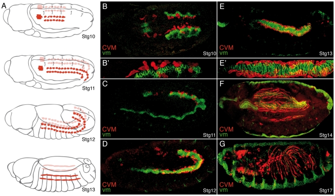
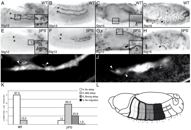
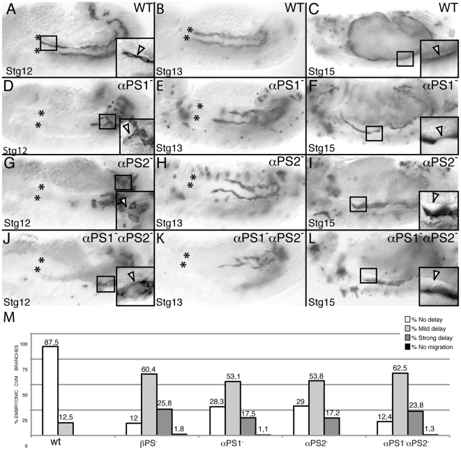
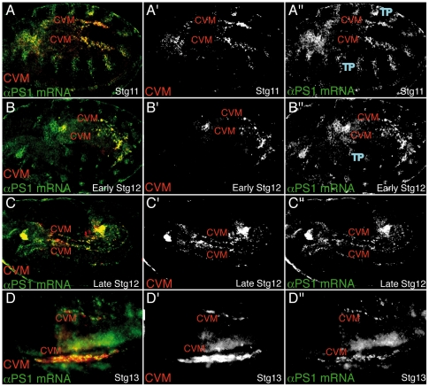
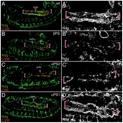
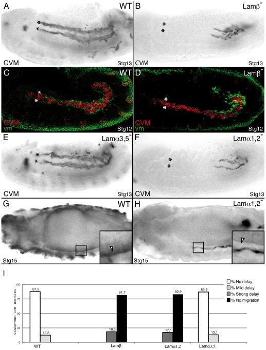
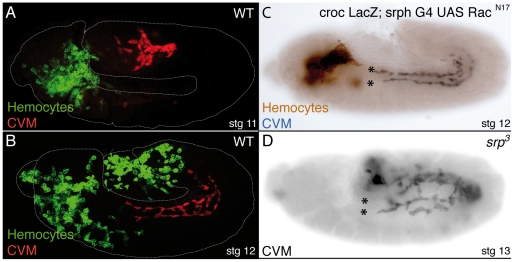
During embryonic development, there are numerous cases where organ or tissue formation depends upon the migration of primordial cells. In the Drosophila embryo, the visceral mesoderm (vm) acts as a substrate for the migration of several cell populations of epithelial origin, including the endoderm, the trachea and the salivary glands. These migratory processes require both integrins and laminins. The current model is that αPS1βPS (PS1) and/or αPS3βPS (PS3) integrins are required in migrating cells, whereas αPS2βPS (PS2) integrin is required in the vm, where it performs an as yet unidentified function. Here, we show that PS1 integrins are also required for the migration over the vm of cells of mesodermal origin, the caudal visceral mesoderm (CVM). These results support a model in which PS1 might have evolved to acquire the migratory function of integrins, irrespective of the origin of the tissue. This integrin function is highly specific and its specificity resides mainly in the extracellular domain. In addition, we have identified the Laminin α1,2 trimer, as the key extracellular matrix (ECM) component regulating CVM migration. Furthermore, we show that, as it is the case in vertebrates, integrins, and specifically PS2, contributes to CVM movement by participating in the correct assembly of the ECM that serves as tracks for migration.


Conflict of interest statement

Competing Interests: The authors have declared that no competing interests exist.

Figures

Figure 1
Figure 1. Migration of the caudal visceral mesodermal cells.
Caudal visceral mesodermal cells (CVM, red) migrate over the visceral mesoderm (vm, green). (A) Schematic diagrams illustrating the two clusters of migrating CVM cells on each side of the embryo (red) at different embryonic stages (adapted from . (B–G) Lateral view, with the exception of B (dorsal view), of embryos carrying the CVM marker croc-LacZ stained for βGal (red) to label CVM cells and FasIII (green) to label the vm. In all figures, embryos are oriented with anterior to the left. (B) At stage (Stg) 10, CVM cells start their migration as two clusters in close contact with the vm (Magnification in B′). (C, D) During Stgs11 and 12, CVM cells move anteriorly over the vm. (E, E′) CVM cells stop migrating at Stg13 when they reach the foregut-midgut transition. (F) At Stg 14, CVM cells spread regularly over the vm. (G) At Stg17, CVM cells have stretched in anterior-posterior direction and form regularly spaced longitudinal fibers.
Figure 2
Figure 2. CVM migration is delayed in embryos lacking the βPS subunit.
(A–D) Wild type embryos and (E–H) βPS maternal and zygotic mutant embryos. CVM cells are visualized by the expression of the transmembrane protein CD2 driven by the CVM G447.2-GAL4 and detected with an anti-CD2 antibody. (A, B, E, F) During germ band retraction, βPS mutant CVM cells show a delay in their migration although they can still send projections as wild type cells do (arrowhead in magnification in black box). (G, H) At Stgs15 and 16, the longitudinal fibers of βPS mutant embryos detach from the underlying vm (G, arrowhead in magnification in black box) and do not spread properly (arrowhead, H). (I, J) Snap shots from live imaging recording embryos carrying the CVM 5053A-GAL4 driving src-GFP. Both wild type (I) and βPS mutant (J) CVM cells send projections (arrowheads) while migrating. (K) Quantification of the CVM migration phenotype in Stg13 embryos of the indicated genotypes. (L) Schematic diagram of a Stg13 embryo showing the distance reached by CVM cells. In all figures, asterisks mark the foregut-midgut transition, where CVM cells stop migrating.
Figure 3
Figure 3. PS1 and PS2 integrins are required for proper CVM migration.
CVM cells are visualized using the combination G447.2-GAL4/UAS-CD2 and an anti-CD2 antibody staining. CVM cells from Stgs12 and 13 αPS1 (D, E, M) and αPS2 (G, H, M) mutant embryos are delayed in their migration as compared with wild type cells (A, B, M, yet they still send projections (D, G, arrowhead in magnification in black box) (F, I) In addition, CVM fibers from these mutant embryos detach from the vm at Stg15 (arrowhead in magnification in black box). (J–L) These phenotypes are enhanced in αPS1αPS2 double mutant embryos, phenocopying the defects observed in βPS mutant embryos. (M) Quantification of the CVM migration phenotype in Stg13 embryos of the designated genotypes.
Figure 4
Figure 4. αPS1 is expressed in migrating CVM cells.
(A–D) Dorso-lateral views of embryos carrying the CVM marker croc-LacZ double-stained with βGal antibody (red) and αPS1 expression by fluorescence in situ hybridization (green). In all panels both clusters of CVM cells (CVM) can be visualized. (A″–D″) αPS1 mRNA is strongly expressed in CVM cells during all phases of their migration, from Stg11 to Stg13.TP, tracheal pits.
Figure 5
Figure 5. αPS1 function is specifically required in CVM cells to mediate their migration.
The ability of different UAS-αPS1 transgenes to rescue the CVM migration defects of Stg 13 αPS1 mutant embryos was assessed by co-expressing UAS-CD2 and the different transgenes using the G447.2-GAL4 and staining with anti-CD2 antibody. (A) Wild type embryo. (B) αPS1 mutant embryo. (C) CVM-specific expression of αPS1, but not αPS2 (D), substantially rescues the CVM migration defects of αPS1 mutant embryos. (E) The αPS1PS2 transgene also shows significant rescue, although less effective than αPS1. (F) Conversely, the αPS2PS1 transgene is as little effective as αPS2. (G) Quantification of the CVM migration phenotype in Stg13 embryos of the indicated genotypes.
Figure 6
Figure 6. PS2 is required for Nidogen accumulation over the vm.
(A–D) Lateral view of Stg13 embryos carrying the CVM marker croc-LacZ double-stained with anti-βGal (red) and anti-Ndg (green) antibodies. (A) In wild type embryos Ndg is found at the edges of vm (pink brackets) along the path of migrating CVM cells. This distribution is affected in βPS (B), αPS2 (C) but not αPS1 (D) mutant embryos.
Figure 7
Figure 7. LamininW, but not lamininA, is required for CVM migration.
CVM cells are visualized using the combination 5053A-GAL4/UAS-srcGFP (A–D, F, G, H) or G447.2-GAL4/UAS-CD2 (E) and anti-GFP or anti-CD2 antibody staining, respectively. (A) Wild type embryo. (B) In absence of Laminin function, CVM cells fail to migrate. (C) During Stg12, wild type CVM cells (red) migrate in close contact with the vm, visualized with anti-FasIII antibody (green). However, Lamβ mutant CVM cells contact an intact vm but fail to migrate (D). (E) CVM migration is unaffected in Lamα3, 5 mutant embryos. (F) Conversely, CVM cells of Lamα1, 2 mutant embryos show a delay in their migration similar to that observed in Lamβ mutant embryos (B). (G, H) Attachment of CVM fibers to the vm is affected in stage 15 Lamα1, 2 mutant embryos (H) compare to wild type (G) (arrowhead in magnification in black box). (I) Quantification of the CVM migration phenotype in Stg13 embryos of the indicated genotypes.
Figure 8
Figure 8. Mature hemocytes and fat body, are not essential for CVM migration.
(A–D) Hemocytes and CVM cells are visualized using the lines srph-GAL4/UAS-CD2 and croc-LacZ, respectively. (A) In wild type Stg11 embryos, CVM cells (red) initiate their migration before hemocytes (green) have populated the posterior end of the embryo. (B) In subsequent stages, CVM migration always precedes that of hemocytes. (C) Blocking hemocyte migration (brown), by expressing RacN17, does not affect CVM migration (black). (D) Absence of mature hemocytes and fat body does not affect CVM migration.

References

    1. Bokel C, Brown NH. Integrins in Development: moving on, respoding to, and sticking to the extracellular matrix. Developmental Cell. 2002;3:311–321. - PubMed
    1. Roote CE, Zusman S. Functions for PS integrins in tissue adhesion, migration, and shape changes during early embryonic development in Drosophila. Dev Biol. 1995;169:322–336. - PubMed
    1. Martin-Bermudo MD, Alvarez-Garcia I, Brown NH. Migration of the drosophila primordial midgut cells requires coordination of diverse PS integrin functions. Development. 1999;126:5161–5169. - PubMed
    1. Boube M, Martin-Bermudo MD, Brown N, Casanova J. Specific tracheal migration is mediated by complementary expression of cell surface proteins. Genes and development. 2001;15:1554–1562. - PMC - PubMed
    1. Devenport D, Brown NH. Morphogenesis in the absence of integrins:mutation of both Drosophila βsubunits prevents midgut migration. Development. 2004;131:5405–5415. - PubMed

Publication types

MeSH terms