. 2011;6(9):e24102.
doi: 10.1371/journal.pone.0024102. Epub 2011 Sep 19.

Efficient elimination of cancer cells by deoxyglucose-ABT-263/737 combination therapy

Affiliations

Efficient elimination of cancer cells by deoxyglucose-ABT-263/737 combination therapy

Ryuji Yamaguchi et al. PLoS One. 2011.

Abstract

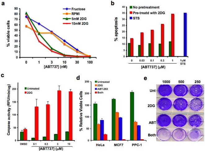
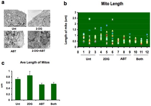
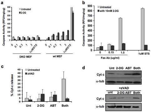
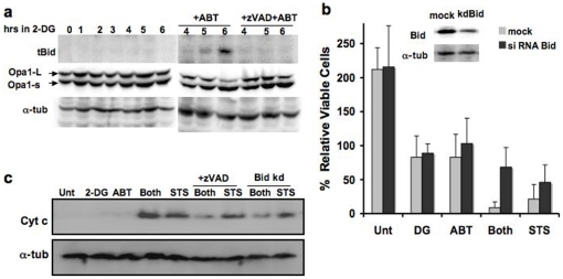
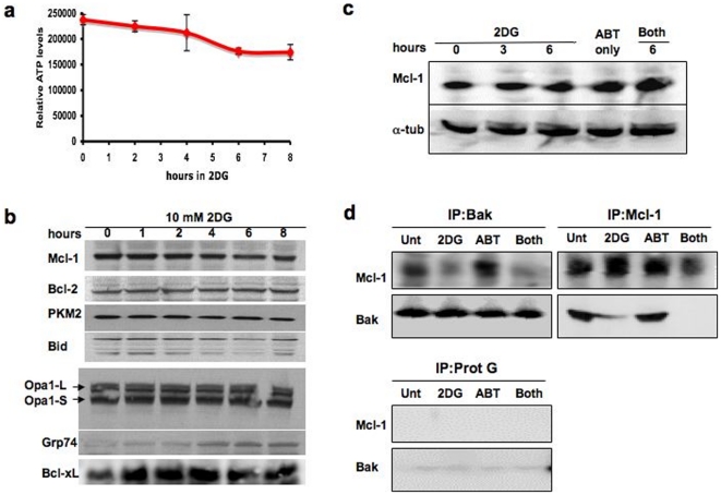
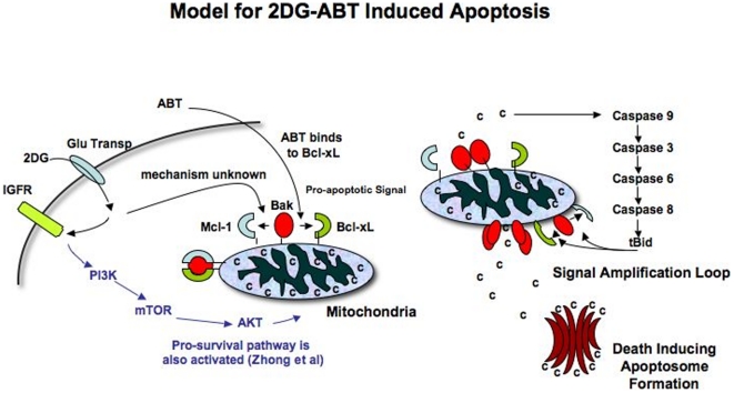
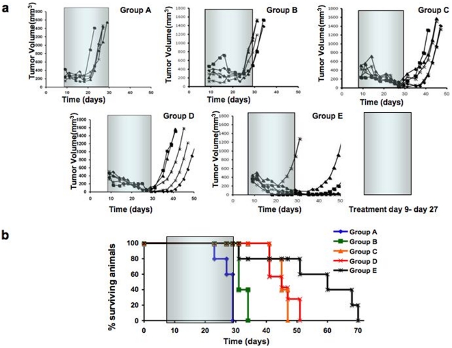
As single agents, ABT-263 and ABT-737 (ABT), molecular antagonists of the Bcl-2 family, bind tightly to Bcl-2, Bcl-xL and Bcl-w, but not to Mcl-1, and induce apoptosis only in limited cell types. The compound 2-deoxyglucose (2DG), in contrast, partially blocks glycolysis, slowing cell growth but rarely causing cell death. Injected into an animal, 2DG accumulates predominantly in tumors but does not harm other tissues. However, when cells that were highly resistant to ABT were pre-treated with 2DG for 3 hours, ABT became a potent inducer of apoptosis, rapidly releasing cytochrome c from the mitochondria and activating caspases at submicromolar concentrations in a Bak/Bax-dependent manner. Bak is normally sequestered in complexes with Mcl-1 and Bcl-xL. 2DG primes cells by interfering with Bak-Mcl-1 association, making it easier for ABT to dissociate Bak from Bcl-xL, freeing Bak to induce apoptosis. A highly active glucose transporter and Bid, as an agent of the mitochondrial apoptotic signal amplification loop, are necessary for efficient apoptosis induction in this system. This combination treatment of cancer-bearing mice was very effective against tumor xenograft from hormone-independent highly metastasized chemo-resistant human prostate cancer cells, suggesting that the combination treatment may provide a safe and effective alternative to genotoxin-based cancer therapies.


Conflict of interest statement

Competing Interests: The authors have declared that no competing interests exist.

Figures

Figure 1
Figure 1. Annexin V-staining of 2DG-ABT treated RS(4;11) and HeLa cells.
a, RS(4;11) cells pre-treated with 20 mM fructose or 0–10 mM 2-deoxy-D-glucose in regular RPMI medium containing 12 mM glucose, for 1 hour before the addition of ABT-737 at indicated concentrations. 9 hours later, cells were analyzed by FACS for Annexin-V positivity. Chi Square Test of Independence for two variables, 2DG and ABT, was P(x~1 2>193.35) = 0.000000. Since the combination was clearly better than expected values, the statistic suggests synergistic interaction between 2DG and ABT-737. b, HeLa cells were pre-treated for 1 hour either with or without 10 mM deoxyglucose in DMEM containing 12 mM glucose, before adding ABT-737. Six hours later, cells were analyzed by FACS for Annexin-V staining. c, HeLa cells were pre-treated for 3 hours with 2DG before the addition of ABT-737. Cells were assayed for caspase activity at 3 hours after ABT-737 addition. d, HeLa, PPC-1 and MCF-7 cells were treated with 10 mM 2DG for 3 hours before adding 1 µM ABT-263 for overnight incubation. Trypan blue-negative (viable) cells were counted and graphed (mean ± std dev; n = 3). e, Combination therapy reduces clonogenic survival of PPC-1 cells. PPC-1 cells were treated just once for 24 hrs with 5 mM 2DG, 1 µM ABT-263, both, or were left untreated. 250–1000 cells were plated per 30 mm dish, and 12 days later, surviving cell colonies were stained.
Figure 2
Figure 2. Combination of 2DG and ABT-263 does not cause mitochondrial outer membrane rupture.
HeLa cells treated with either 10 mM 2DG for 6 hours, 1 µM ABT-263 for 3 hours, or 2DG for 3 hours before adding ABT-263 for an additional 3 hours, or left untreated. a, Cells were fixed for electron microscopy (see Method). Three cells from each treatment group were examined closely for the length of mitochondria. We did not observe a single instance of ruptured mitochondrial outermembranes. b, From each cell, a minimum of 12 mitochondria were randomly chosen for measurement. c, Average lengths of mitochondria became shorter with treatments containing ABT-263 (mean +/− std dev ). Since ABT-263/737 binds to Bcl-2 family members, which are known to be involved in mitochondrial fusion/fission, ABT may have altered the balance of mitochondrial fusion/fission to the fission side (p = 0.0001, 95% confidence interval of this difference: from 14.5407 to 37.2513; unpaired t-test).
Figure 3
Figure 3. 2DG-ABT induced apoptosis is a non-canonical mitochondria-dependent form of apoptosis.
a, Wt, and Bax/Bak DKO MEFs were treated with 10 mM 2DG for 3 hours before the addition of 3 µM ABT-263 for 3 hours. 3 hours later, cells were harvested for caspase activity assays. (See also Fig. S5a). b, HT1080 cells were treated with or without 2DG for 3 hours. Then cells were treated with either 1 µM STS or anti-Fas (CD95) antibody at indicated concentrations (µg/ml). Cells were harvested 3 hours after, and assayed for caspase activity. Interestingly, 2DG seems to have interfered with Fas-induced apoptosis. In repeated experiments, we observed consistently lower rates of caspase activation in 2DG pre-treated cells, but the degrees of inhibitory activity varied from experiment to experiment. We are not sure of its cause. c, For the first 3 hours, HeLa cells were either treated with 10 mM 2DG or left untreated and then treated with 10 µM z-VAD, 1 µM ABT-737, both, or neither for 3 more hours. The cells were fixed and stained for DNA (red) and cytochrome c (green) and examined for loss of cytochrome c staining, as described in Methods. Bar indicates 100 µm. The representative images are shown in Fig. S3. This graph shows the percentage of cells with loss of cytochrome c staining. The combination treatment had higher rates of cells with released cytochrome c (p = 0.008 by unpaired t-test ). d, Cells treated as in c were fractionated and cytosolic fractions were analyzed by western blots.
Figure 4
Figure 4. The Bid-based feedback loop is required for efficient 2DG-ABT induced apoptosis.
a, Bid is activated during 2DG-ABT induced apoptosis. Western blots of PPC-1 cell lysates treated with 10 mM 2DG for 0–6 hours (left panel), with either 1 µM ABT-263 alone or ABT-263+10 µM z-VAD for the last 3 hours (right panel), probed with the anti-tBid antibody (upper panels), anti-Opa1 antibody (middle panels), and anti-alpha-tubulins (lower panels). Note: the loss of Opa-1-L was used as a marker for remodeled mitochondrial cristae during cytochrome c release from the mitochondria . b, The effects of Bid depletion on 2DG-AB-induced apoptosis. Insert: western blots of PPC-1 cell lysates, mock transfected (left lane) or transfected with Bid siRNA (right lane). Bid (+) and Bid (−) PPC-1 cells were treated with either 2DG, ABT-263 (1 µM), both, 10 µM ABT-263, or left untreated for 24 hours. The viable cells were counted and the increase/decrease over the input numbers were graphed. c, In PPC-1 cells either pre-treated with 2DG for 3 hours or without pre-treatment, then 1 µM ABT-263 or 1 µM STS was added for three hours, and cytosolic fractions were analyzed by western blots. In parallel experiments, Bid-depleted PPC-1 cells were treated with the combination of 2DG and ABT-263 or with STS and analyzed (designated as Bid kd, last 2 lanes). Cells were harvested 3 hour after ABT/STS treatment, and cytosolic fractions were analyzed with western blots.
Figure 5
Figure 5. 2DG disrupts Bak-Mcl-1 association, priming cancer cells without lowering protein levels of Mcl-1.
a, The effect of 2DG on cellular ATP levels. The amounts of ATP were measured during HeLa cell culture with 10 mM 2DG in the presence of 25 mM glucose in DMEM supplemented with 10% serum. b, Protein profiles during 2DG incubation of HeLa cells. Samples were taken from HeLa cells treated as in a, and various protein contents of pro- and anti-apoptotic proteins were analyzed by Western blots. In this experiment, we see increases in Bcl-xL for the first hour. When we repeated the experiment twice, Bcl-xL levels remained the same. c, Mcl-1 levels remained unchanged during 2DG-ABT-induced apoptosis. PPC-1 cells were treated with 10 mM 2DG for 6 hours, 1 µM ABT-263 for 3 hours, or 10 mM 2DG for 6 hours of which the last 3 hours were incubated with 1 µM ABT-263. d, Bak and Mcl-1 do not co-precipitate in 2DG treated cells. PPC-1 cells were treated as in c. Bak and Mcl-1 were immunoprecipitated by specific antibodies conjugated to Protein G-Sepharose and the co-precipitated proteins were analyzed by Western blots. Left Upper Panels, Bak co-precipitates, Right Upper Panels, Mcl-1 co-precipitates, and Left Lower Panels, Protein G-Sepharose precipitates. For technical reasons, we could not immunoprecipitate Bcl-xL and analyze its content.
Figure 6
Figure 6. Model of 2DG-ABT Induced Apoptosis.
a, 2DG is specifically taken up by cancer cells through a glucose transporter. Once inside, it is phosphorylated and becomes a hexokinase inhibitor. At the same time, it activates several signal transduction cascades. One of them is IGFR-PI3K-mTOR-AKT pro-survival pathway. Through another cascade activation, Bak-Mcl-1 association is lost. When ABT is added 3 hours later, it can bind to Bcl-xL and disrupts its association with Bak. b, Freed Bak oligomerizes, releasing a small amount of cytochrome c into the cytosol. It activates cascades of caspases 9-3-6-8 and caspase 8 cleaves Bid, generating BH3 only tBid protein. Since tBid can dissociate Bak-Mcl-1 and Bak-Bcl-xL association, it induces mitochondria outermembrane pore formation through which more cytochrome can be released. Released cytochrome c recruits Apaf1, caspase 9 and dATP, forming a “death executioner” apotosome complex.
Figure 7
Figure 7. Treatment of tumor-bearing mice with 2DG-ABT combination.
Five million PPC-1 human prostate cancer cells were xenografted onto nude mice as described in the text and the method. a, Mice Tumor volumes were measured in mice (n = 7 per group) treated with diluent only (group A), 2DG (group B), ABT-263 (group C), 2DG plus ABT-263 (group D), or 2DG mixed with D-glucose plus ABT-263 (group E), as explained in the text, where drugs were administered 3 times weekly between day 9 and 27 (gray rectangles). b, Survival of tumor-bearing mice was determined at various times post-inoculation with PPC-1 cells. The % of surviving mice is indicated from groups A-E (n = 7 per group). The difference of survival days between Group C & D versus Group E is p = 0.0283, 95% confidence interval of this difference: from −20.98 to −1.36 by unpaired t-test.

Similar articles

Cited by

References

    1. Pelicano H, Martin DS, Xu RH, Huang P. Glycolysis inhibition for anticancer treatment. Oncogene. 2006;25:4633–4646. - PubMed
    1. Caraco C, Aloj L, Chen LY, Chou JY, Eckelman WC. Cellular release of [18F]2-fluoro-2-deoxyglucose as a function of the glucose-6-phosphatase enzyme system. J Biol Chem. 2000;275:18489–18494. - PubMed
    1. Maschek G, Savaraj N, Priebe W, Braunschweiger P, Hamilton K, et al. 2-deoxy-D-glucose increases the efficacy of adriamycin and paclitaxel in human osteosarcoma and non-small cell lung cancers in vivo. Cancer Res. 2004;64:31–34. - PubMed
    1. Dearling JL, Qureshi U, Begent RH, Pedley RB. Combining radioimmunotherapy with antihypoxia therapy 2-deoxy-D-glucose results in reduction of therapeutic efficacy. Clin Cancer Res. 2007;13:1903–1910. - PubMed
    1. Zhong D, Liu X, Schafer-Hales K, Marcus AI, Khuri FR, et al. 2-Deoxyglucose induces Akt phosphorylation via a mechanism independent of LKB1/AMP-activated protein kinase signaling activation or glycolysis inhibition. Mol Cancer Ther. 2008;7:809–817. - PubMed

Publication types

MeSH terms