. 2011;6(9):e24353.
doi: 10.1371/journal.pone.0024353. Epub 2011 Sep 16.

Pathogenic Neisseria hitchhike on the uropod of human neutrophils

Affiliations

Pathogenic Neisseria hitchhike on the uropod of human neutrophils

Niklas Söderholm et al. PLoS One. 2011.

Abstract

Polymorphonuclear neutrophils (PMNs) are important components of the human innate immune system and are rapidly recruited at the site of bacterial infection. Despite the effective phagocytic activity of PMNs, Neisseria gonorrhoeae infections are characterized by high survival within PMNs. We reveal a novel type IV pilus-mediated adherence of pathogenic Neisseria to the uropod (the rear) of polarized PMNs. The direct pilus-uropod interaction was visualized by scanning electron microscopy and total internal reflection fluorescence (TIRF) microscopy. We showed that N. meningitidis adhesion to the PMN uropod depended on both pilus-associated proteins PilC1 and PilC2, while N. gonorrhoeae adhesion did not. Bacterial adhesion elicited accumulation of the complement regulator CD46, but not I-domain-containing integrins, beneath the adherent bacterial microcolony. Electrographs and live-cell imaging of PMNs suggested that bacterial adherence to the uropod is followed by internalization into PMNs via the uropod. We also present data showing that pathogenic Neisseria can hitchhike on PMNs to hide from their phagocytic activity as well as to facilitate the spread of the pathogen through the epithelial cell layer.


Conflict of interest statement

Competing Interests: The authors have declared that no competing interests exist.

Figures

Figure 1
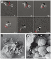
Figure 1. Neisseria adhere to human PMNs.
(A) Image sequences showing the interaction between a bacterial microcolony of N. gonorrhoeae and a PMN. The bacterial microcolony escapes engulfment and move along the plasma membrane to the uropod. Images show 120 seconds of live-cell imaging. Red lines indicate the pseudopods and the front of the migrating PMN. The white ring indicates the microcolony of interest. (B) SEM electrograph showing pilus-mediated adherence of N. meningitidis FAM20 to the plasma membrane on the right side of the pseudopod. (C) Magnified image of (B). Arrows indicate pili.
Figure 2
Figure 2. Bacterial microcolonies adhere to the uropod.
(A) DIC images of viable PMNs with N. meningitidis FAM20 adhered to the uropod. Three images show; low bacterial adherence (left), a medium-sized microcolony (middle) and a big microcolony adhering to the uropod (right). Red lines indicate the pseudopods and the front of the migrating PMN. (B) SEM electrograph showing the specificity of FAM20 adherence to the uropod of a polarized PMN. (C) TEM image showing the interaction between N. gonorrhoeae MS11 P+ and a PMN. Bacterial uropod adherence is enlarged in the box to the right.
Figure 3
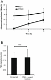
Figure 3. Bacterial adherence occurs frequently and does not impair PMN velocity.
(A) The frequency of bacterial adherence to a PMN uropod. Graph showing the number of PMNs with bacteria adhering to the uropod after 1 to 3 hours of incubation. The adherence of MS11 and FAM20 to the uropod of PMNs was calculated from more than 300 observed cells in three independent experiments. The average percentages of adhering N. gonorrhoeae MS11 and N. meningitidis FAM20 and standard deviations are shown. (B) Graph showing the average velocity of PMNs migrating on glass. Differences in average velocity were determined by Student t-test (p<0.05). The mean velocity of uninfected and infected PMNs and standard deviations are shown.
Figure 4
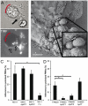
Figure 4. Pili promote adherence of Neisseria to the uropod and meningococcal adherence depends on PilC expression.
(A) The interaction between DyLight 488 NHS ester-stained pili and the PMNs was observed under the microscope in TIRF using a connected argon laser and a 100x objective (N/A 1.46). Red lines indicate the pseudopod. Upper panel: the PMN in bright field image. Arrow indicates one adhering microcolony of FAM20 bacteria. Lower panel: TIRF image of the PMN. White arrows indicate two pili bound to the plasma membrane of the uropod. (B) SEM electrograph showing the direct pilus-uropod interaction. (C) Graph showing the average number of PMNs with N. gonorrhoeae MS11 and mutants adhering to the uropod. (D) Graph showing the average number of PMNs with N. meningitidis FAM20 and mutants adhering to the uropod. Bacterial adherence was calculated from more than 300 observed cells in three independent experiments. One hundred cells were observed with the FAM20 ΔpilT. Statistical significance was calculated in C and D by one-way ANOVA. Data were considered significant if P<0.05 and indicated as *.
Figure 5
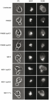
Figure 5. CD46 accumulates at the uropod adjacent to adhering bacteria.
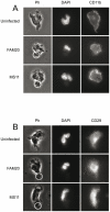
N. gonorrhoeae MS11 and N. meningitidis FAM20 bacteria were allowed to adhere to freshly isolated PMNs. Cells were fixed, permeabilized and incubated with polyclonal antibodies against CD46 and cellular DNA was stained with DAPI. Phase contrast images show the PMN morphology and bacterial adherence. Circles indicate bacteria. Representative images of CD46 expression in PMNs infected with wild-type and mutant gonococci and meningococci are shown.
Figure 6
Figure 6. CD11b and CD29 expression in infected and uninfected PMNs.
N. gonorrhoeae MS11 and N. meningitidis FAM20 bacteria were allowed to adhere to freshly isolated PMNs. Cells were fixed, permeabilized and incubated with monoclonal antibodies against CD11b or CD29 and cellular DNA was stained with DAPI. Phase contrast images show the PMN morphology and bacterial adherence. Circles indicate bacteria. (A) Representative images of CD11b expression in PMNs infected with wild-type and mutant gonococci and meningococci. (B) Representative images of CD29 expression in PMNs infected with wild-type and mutant gonococci and meningococci.
Figure 7
Figure 7. Bacteria invade PMNs via the uropod and intracellular bacteria can be found close to the uropod.
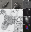
(A) N meningitidis FAM20 were allowed to adhere to the uropod of freshly isolated PMNs. Left electron micrograph shows intracellular bacteria at the uropod. Right electron micrograph shows a magnified image of the cellular uropod at 16 000 x magnification of the same PMN. Arrow indicates an intracellular bacterium covered by the plasma membrane. (B) TEM image showing two cellular compartments in which intracellular bacteria reside. Upper enlarged box shows a bacterium inside a phagosome and the lower enlarged box shows a bacterium in a different compartment situated at the uropod. (C) DyLight NHS ester-stained N meningitidis FAM20 were allowed to adhere to the uropod of freshly isolated PMNs. Cells were stained with fluorescent labeled Lysotracker. An image of an infected PMN (phase contrast), DyLight NHS ester-stained intracellular meningococci (green), and acidic compartments (red) is shown. Arrows indicate bacteria.
Figure 8
Figure 8. PMN interacts with bacteria bound to epithelial cells.
N. meningitidis FAM20 were allowed to form microcolonies and adhere to FaDu cells. After one hour, freshly isolated PMNs were added. Representative DIC image sequences at 63x magnification from at least three independent live-cell time-lapse experiments are shown. Red lines indicate the pseudopods and the front of the migrating PMN. (A) PMN migrating towards microcolonies but is immobilized and trapped by a microcolony that is bound to both an epithelial cell and a uropod. (B) The PMN removes the microcolony from the epithelial cell and transports it away from the site of initial adherence. The dotted black line indicates the borderline of the FaDu cells. (C) A microcolony (green) adheres to the PMN (blue) and penetrates a cell layer by hitchhiking on a PMN. The red lines indicate PMN pseudopods and the front of the migrating PMN. Images were processed by Adobe Photoshop.
Figure 9
Figure 9. A schematic illustration of bacteria hitchhiking on PMNs.
(A) Bacterial adherence to PMNs occurs either by direct uropod contact or by transport on the plasma membrane from the pseudopod. Although initial contact was made with the pseudopod, PMNs ended up bound to the microcolony via their uropod. The binding recruits CD46 and bacteria can be internalized via the uropod. (B) The PMN can be immobilized and trapped by a microcolony that is bound to both an epithelial cell and a uropod or (C) the PMN can remove the microcolony from the epithelial cell and transport it away from the site. (D) The nature of PMNs allows them to easily migrate under and in between epithelial cells. In this manner, bacteria can hitchhike on the uropod to penetrate the epithelial barrier.

Similar articles

Cited by

References

    1. Nassif X, So M. Interaction of pathogenic neisseriae with nonphagocytic cells. Clin Microbiol Rev. 1995;8:376–388. - PMC - PubMed
    1. Coureuil M, Lecuyer H, Scott MG, Boularan C, Enslen H, et al. Meningococcus Hijacks a beta2-adrenoceptor/beta-Arrestin pathway to cross brain microvasculature endothelium. Cell. 2010;143:1149–1160. - PubMed
    1. Sjolinder H, Jonsson AB. Olfactory nerve–a novel invasion route of Neisseria meningitidis to reach the meninges. PLoS One. 2010;5:e14034. - PMC - PubMed
    1. Kellogg DS, Cohen IR, Norins LC, Schroeter AL, Reising G. Neisseria gonorrhoeae. II. Colonial variation and pathogenicity during 35 months in vitro. J Bacteriol. 1968;96:596–605. - PMC - PubMed
    1. Swanson J. Studies on gonococcus infection. IV. Pili: their role in attachment of gonococci to tissue culture cells. J Exp Med. 1973;137:571–589. - PMC - PubMed

Publication types

Substances