. 2011;6(9):e24660.
doi: 10.1371/journal.pone.0024660. Epub 2011 Sep 19.

HCV causes chronic endoplasmic reticulum stress leading to adaptation and interference with the unfolded protein response

Affiliations

HCV causes chronic endoplasmic reticulum stress leading to adaptation and interference with the unfolded protein response

Emmanuelle Merquiol et al. PLoS One. 2011.

Abstract

Background: The endoplasmic reticulum (ER) is the cellular site for protein folding. ER stress occurs when protein folding capacity is exceeded. This stress induces a cyto-protective signaling cascades termed the unfolded protein response (UPR) aimed at restoring homeostasis. While acute ER stress is lethal, chronic sub-lethal ER stress causes cells to adapt by attenuation of UPR activation. Hepatitis C virus (HCV), a major human pathogen, was shown to cause ER stress, however it is unclear whether HCV induces chronic ER stress, and if so whether adaptation mechanisms are initiated. We wanted to characterize the kinetics of HCV-induced ER stress during infection and assess adaptation mechanisms and their significance.

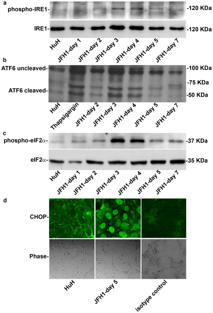
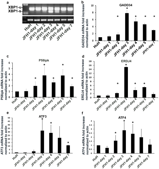
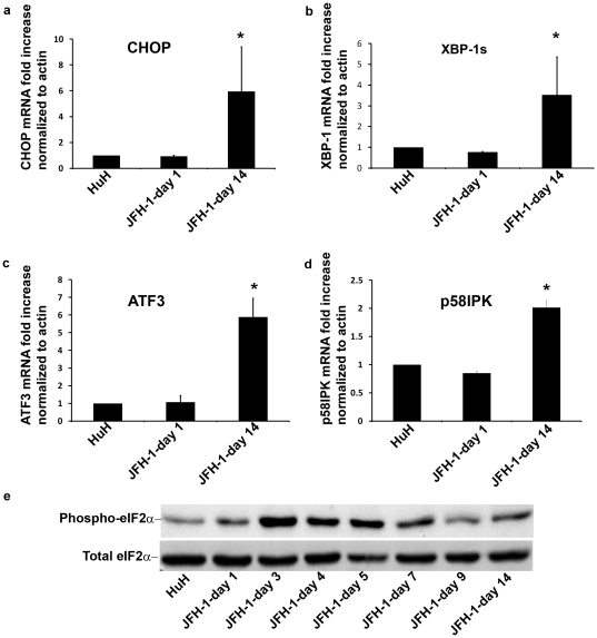
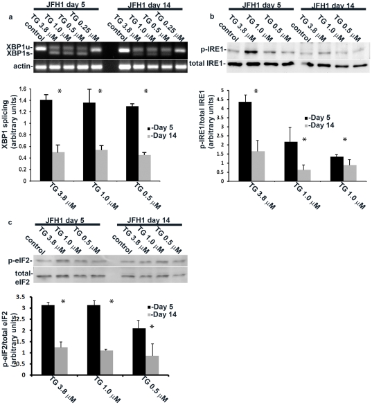
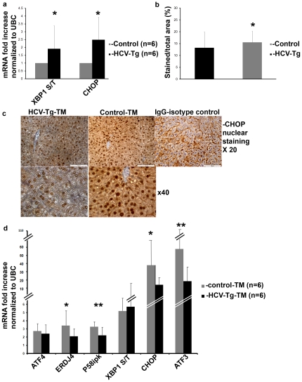
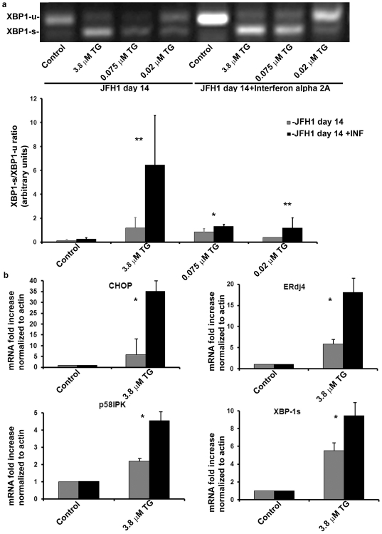
Methods and findings: The HuH7.5.1 cellular system and HCV-transgenic (HCV-Tg) mice were used to characterize HCV-induced ER stress/UPR pathway activation and adaptation. HCV induced a wave of acute ER stress peaking 2-5 days post-infection, which rapidly subsided thereafter. UPR pathways were activated including IRE1 and EIF2α phosphorylation, ATF6 cleavage and XBP-1 splicing. Downstream target genes including GADD34, ERdj4, p58ipk, ATF3 and ATF4 were upregulated. CHOP, a UPR regulated protein was activated and translocated to the nucleus. Remarkably, UPR activity did not return to baseline but remained elevated for up to 14 days post infection suggesting that chronic ER stress is induced. At this time, cells adapted to ER stress and were less responsive to further drug-induced ER stress. Similar results were obtained in HCV-Tg mice. Suppression of HCV by Interferon-α 2a treatment, restored UPR responsiveness to ER stress tolerant cells.

Conclusions: Our study shows, for the first time, that HCV induces adaptation to chronic ER stress which was reversed upon viral suppression. These finding represent a novel viral mechanism to manipulate cellular response pathways.


Conflict of interest statement

Competing Interests: The authors have declared that no competing interests exist.

Figures

Figure 1
Figure 1. HCV infection induces a wave like activation of the 3 arms of the UPR.
HuH7.5.1 hepatoma cells were infected with HCV-JFH1. Following infection, protein was extracted at the indicated time points. The expression of (a) phospho-IRE1, and (b) cleaved and non-cleavedATF6 (c) phospho-eIF2α were analyzed by western blotting. (d) CHOP translocation to the nucleus was assessed by immunofluorecence in control HuH7.5.1 cells or JFH1 infected cells on day 5, isotype anti-rabbit IgG was used as control. Results are means±S.D. of 3 experiments. Blots and picture are from a representative experiment (see also Figure S3a).
Figure 2
Figure 2. Downstream target genes of the UPR are up-regulated following HCV infection.
HuH7.5.1 hepatoma cells were infected with HCV-JFH1. Following infection, cells were harvested and RNA was extracted at the indicated time points. (a) splicing of XBP-1 was measured by RT-PCR. Gene expression levels of (b) GADD34 (c) p58ipk (d) ERDJ4 (e) ATF3 and (f) ATF4 were determined by real-time PCR relative to b-actin. Results are means±S.D. of 3 experiments. * = p<0.05 (ANOVA, one way) versus non-infected cells.
Figure 3
Figure 3. HCV infection induces prolonged activation of the UPR.
HCV infected HuH7.5.1 cells were grown in culture for 14 days and compared to non-infected cells and day 1 post-infection cells. Cells were harvested, RNA extracted and gene expression levels were determined by real-time PCR and normalized to b-actin expression. Results are means±S.D. of 3 independent experiments. * = p<0.05 (ANOVA, one way) for day 14 versus non-infected and day 1 infected cells.
Figure 4
Figure 4. HCV-induced chronic ER stress confers resistance to drug induced UPR.
HCV infected cells were stimulated by decreasing doses of thapsigargin at day 5 and at day 14 post infection and the effect of stimulation on (a) XBP-1 splicing or (b) IRE1 and (c) eIF2α phosphorylation was assessed by RT-PCR and immunoblotting respectively. Quantification of the results was obtained from densitometric scanning of the blots (means±S.D.) of 3 experiments. * = p<0.05 (ANOVA, one way) for day 5 versus day 14 post infection.
Figure 5
Figure 5. HCV-Tg mice display chronic ER stress and activate UPR genes less efficiently than controls following ER stress induction.
Untreated HCV-Tg and control mice were sacrificed, liver excised and RNA was extracted. (a) Expression of the ratio of XBP-1s to XBP-1u (S/T) and CHOP were assessed by real-time PCR. (Panels b, c and d) Mice were treated i.p. with tunicamycin injection of 1 mg/kg every other day (total of 2–3 injections), and were sacrificed 3 days following the last injection. (c) Immunostaining of formalin-fixed livers for CHOP (b) Quantification of stained nuclei was done automatically by visualizing the slides on an Olympus BX61 microscope, photography was performed on Photometric CoolSnap ES and analysis was done using Ariol SL-50 Applied Imaging 3.1.270 software. Untreated mice received 5% dextrose injection.(d) gene expression levels were determined by real-time PCR relative to UBC expression. Results are means±S.D. of 3 independent experiments. *p<0.05, **p<0.01 (ANOVA, one way) for (a) untreated control versus HCV-Tg mice and for (b, d) HCV-Tg control versus HCV-Tg tunicamycin treated mice.
Figure 6
Figure 6. Interferon α treatment reverses the adaptation to chronic ER stress.
HCV infected cells were treated with Interferon á 2a (1800 IU/ml were added to cell growth medium and changed daily), or left untreated. On day 14 post infection, cells were stimulated with thapsigargin as indicated. (a) XBP-1 splicing was assessed by RT-PCR. Quantification of the results was obtained from densitometric scanning of the blots (means±S.D.) of 3 experiments. * = p<0.05, **p<0.01 for interferon treated versus non-treated cells. (b) Gene expression levels were determined by real-time PCR and normalized to b-actin expression. Results are means±S.D. of 3 independent experiments. *p<0.05, (ANOVA, one way) for interferon treated versus non-treated cells.

References

    1. Shepard CW, Finelli L, Alter MJ. Global epidemiology of hepatitis C virus infection. Lancet Infect Dis. 2005;5(9):558–67. - PubMed
    1. Gale M, Jr, Foy EM. Evasion of intracellular host defence by hepatitis C virus. Nature. 2005;436(7053):939–45. - PubMed
    1. Pereira AA, Jacobson IM. New and experimental therapies for HCV. Nat Rev Gastroenterol Hepatol. 2009;6(7):403–11. - PubMed
    1. Dubuisson J, Penin F, Moradpour D. Interaction of hepatitis C virus proteins with host cell membranes and lipids. Trends Cell Biol. 2002;12(11):517–23. - PubMed
    1. Okamoto K, Moriishi K, Miyamura T, Matsuura Y. Intramembrane proteolysis and endoplasmic reticulum retention of hepatitis C virus core protein. J Virol. 2004;78(12):6370–80. - PMC - PubMed

Publication types

MeSH terms

Substances