. 2011;6(9):e24790.
doi: 10.1371/journal.pone.0024790. Epub 2011 Sep 16.

Structure and molecular evolution of CDGSH iron-sulfur domains

Affiliations

Structure and molecular evolution of CDGSH iron-sulfur domains

Jinzhong Lin et al. PLoS One. 2011.

Abstract

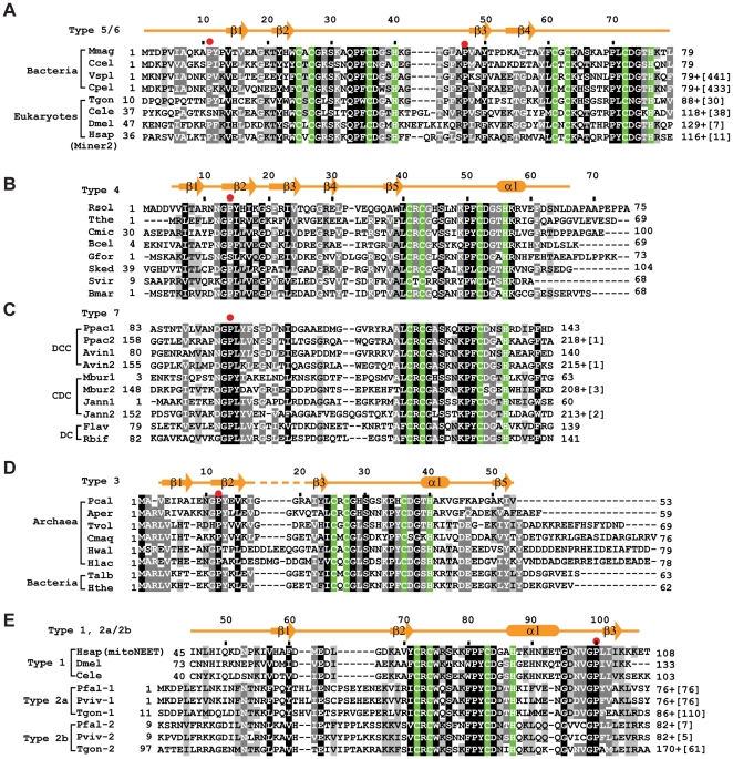
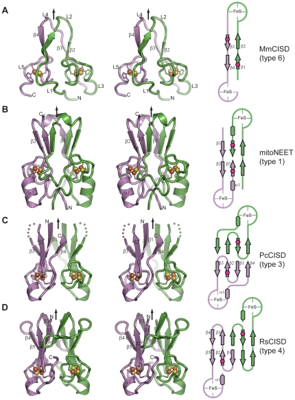
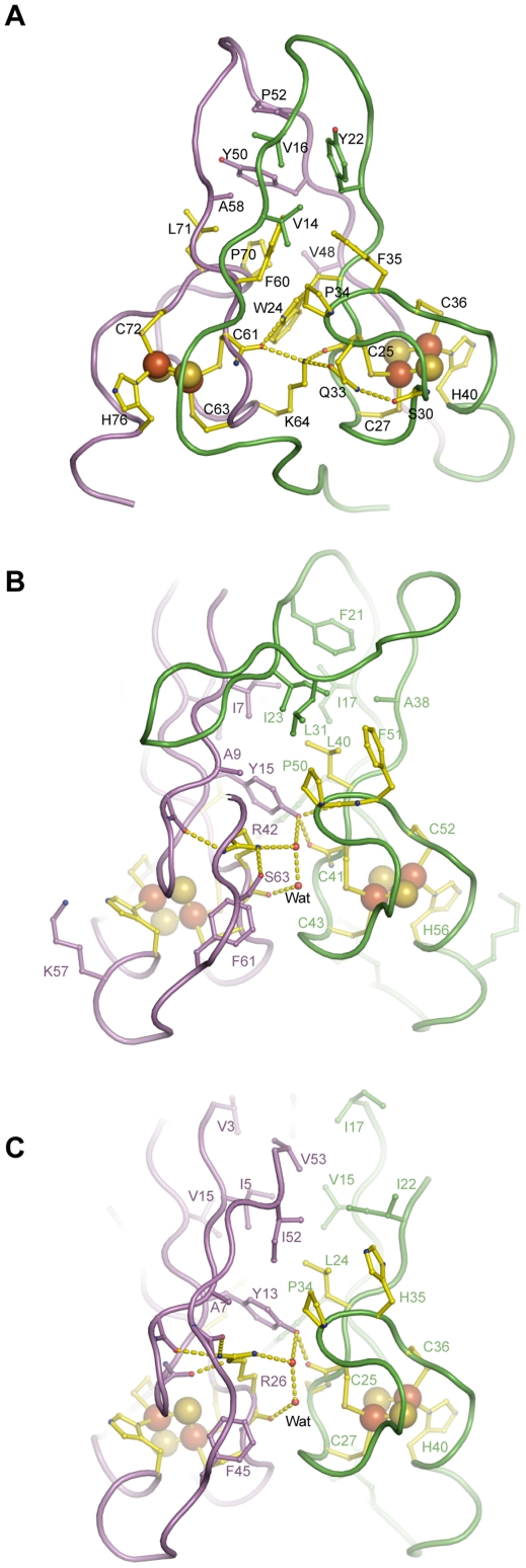
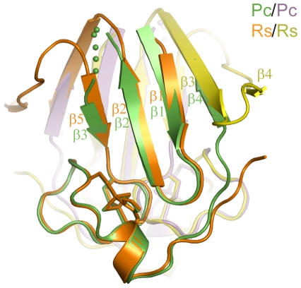
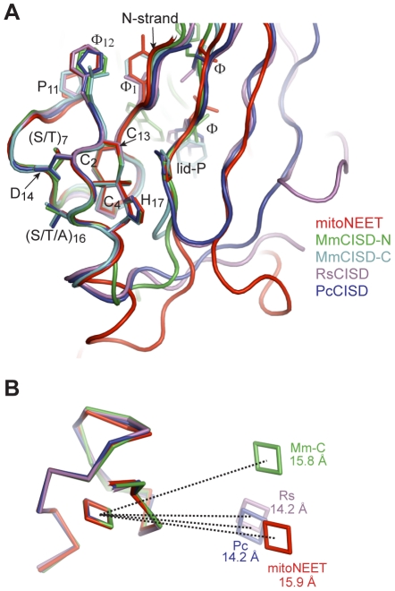
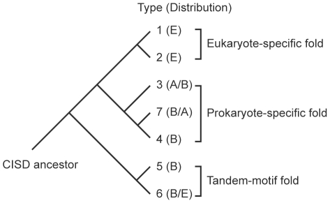
The recently discovered CDGSH iron-sulfur domains (CISDs) are classified into seven major types with a wide distribution throughout the three domains of life. The type 1 protein mitoNEET has been shown to fold into a dimer with the signature CDGSH motif binding to a [2Fe-2S] cluster. However, the structures of all other types of CISDs were unknown. Here we report the crystal structures of type 3, 4, and 6 CISDs determined at 1.5 Å, 1.8 Å and 1.15 Å resolution, respectively. The type 3 and 4 CISD each contain one CDGSH motif and adopt a dimeric structure. Although similar to each other, the two structures have permutated topologies, and both are distinct from the type 1 structure. The type 6 CISD contains tandem CDGSH motifs and adopts a monomeric structure with an internal pseudo dyad symmetry. All currently known CISD structures share dual iron-sulfur binding modules and a β-sandwich for either intermolecular or intramolecular dimerization. The iron-sulfur binding module, the β-strand N-terminal to the module and a proline motif are conserved among different type structures, but the dimerization module and the interface and orientation between the two iron-sulfur binding modules are divergent. Sequence analysis further shows resemblance between CISD types 4 and 7 and between 1 and 2. Our findings suggest that all CISDs share common ancestry and diverged into three primary folds with a characteristic phylogenetic distribution: a eukaryote-specific fold adopted by types 1 and 2 proteins, a prokaryote-specific fold adopted by types 3, 4 and 7 proteins, and a tandem-motif fold adopted by types 5 and 6 proteins. Our comprehensive structural, sequential and phylogenetic analysis provides significant insight into the assembly principles and evolutionary relationship of CISDs.


Conflict of interest statement

Competing Interests: The authors have declared that no competing interests exist.

Figures

Figure 1
Figure 1. Sequence alignment of CISDs.
The secondary structure elements, if available, are shown for the first protein in each alignment. The four residues coordinating the [2Fe-2S] cluster are shaded in green. Residues conserved in 100%, 80% and 60% of the displayed sequences are shaded in black, gray and light gray, respectively. The full lists of aligned CISDs are supplied as fasta files in the Supporting Information. The numbers of starting and ending residues of each sequence are labeled and the numbers of extra residues at the C-terminus are shown in brackets. Red dots indicate the lid-proline. (A) Alignment of types 5 and 6 CISDs. Mmag: Magnetospirillum magneticum, YP_423370 (Genebank ID); Ccel: Clostridium cellulolyticum, YP_002506010; Vspl: Vibrio splendidus, ZP_00989737; Cpel: Candidatus Pelagibacter ubique, YP_265568; Tgon: Toxoplasma gondii, XP_002369458; Cele: Caenorhabditis elegans, NP_497419; Dmel: Drosophila melanogaster, NP_610234; Hsap: Homo sapiens, NP_001129970. Vspl and Cpel are of type 5 and the others are of type 6. (B) Alignment of type 4 CISDs. Rsol: Ralstonia solanacearum, NP_521033; Tthe: Thermus thermophilus, YP_143292; Cmic: Clavibacter michiganensis, YP_001222006; Bcel: Bacteroides cellulosilyticus, ZP_03679950; Gfor: Gramella forsetii, YP_861977; Sked: Sanguibacter keddieii, YP_003314086; Svir: Streptomyces viridochromogenes, ZP_05536111; Bmar: Blastopirellula marina, ZP_01088744. (C) Alignment of type 7 CISDs. Different subtypes DCC, CDC and DC are indicated. The numbers 1 and 2 in the protein name indicate the first and second CISD in subtypes DCC and CDC. Ppac: Plesiocystis pacifica, ZP_01908188; Avin: Allochromatium vinosum, YP_003442661; Mbur: Methanococcoides burtonii, YP_566882; Jann: Jannaschia sp. CCS1, YP_509212; Flav: Flavobacteriales bacterium, ZP_02182054; Rbif: Robiginitalea biformata, YP_003195727. (D) Alignment of type 3 CISDs. Pcal: Pyrobaculum calidifontis, YP_001056297; Aper: Aeropyrum pernix, NP_148024; Tvol: Thermoplasma volcanium, NP_110689; Cmaq: Caldivirga maquilingensis, YP_001540894; Hwal: Haloquadratum walsbyi, YP_658160; Hlac: Halorubrum lacusprofundi, YP_002565978; Talb: Thermocrinis albus, YP_003472998; Hthe: Hydrogenobacter thermophilus, YP_003433409. (E) Alignment of type 1, 2a and 2b CISDs. Hsap, Homo sapiens, NP_060934; Dmel, Drosophila melanogaster, NP_651684; Cele: Caenorhabditis elegans, NP_001022387; Pfal-1 and Pfal-2: Plasmodium falciparum 3D7, XP_001351102 and XP_002808656; Pviv-1 and Pviv-2: Plasmodium vivax SaI-1, XP_001613049 and XP_001615077; Tgon-1 and Tgon-2: Toxoplasma gondii ME49, XP_002365250 and XP_002369458.
Figure 2
Figure 2. Crystal structure and topology schematic of CISDs.
(A–D) Structure and topology of type 6 MmCISD (A), type 1 mitoNEET (PDB: 2QD0 [6]) (B), type 3 PcCISD (C), and type 4 RsCISD (D). Cross-eye stereo views are shown. The two subunits or two halves for the MmCISD structure are colored in green and violet. The secondary structural elements and the N- , C-terminus are indicated for one subunit. Arrows denote the dyad axis or pseudo dyad axis for MmCISD. Red dots in schematic represent the lid-proline.
Figure 3
Figure 3. Dimer interface of CISDs.
(A) The interface between the two halves of the MmCISD structure. (B) The dimer interface of the RsCISD structure. (C) The dimer interface of the PcCISD structure. Key residues are shown as sticks and balls. Oxygen atoms are red, nitrogen atoms are blue, carbon atoms in the CDGSH motif are yellow, and the remaining carbon atoms in one subunit or half are violet and those in the other subunit or half are green. Hydrogen bonds are shown as dashed lines. Only half of the interactions are displayed for the symmetric RsCISD and PcCISD dimer structures.
Figure 4
Figure 4. Alignment of RsCISD and PcCISD structures.
Both subunits are aligned. The two subunits of PcCISD are colored in green and purple, and the two subunits of RsCISD are colored in orange and yellow.
Figure 5
Figure 5. Structural conservation of CISDs.
(A) Superimposition of the five nonequivalent iron-sulfur binding modules from RsCISD, MmCISD PcCISD and mitoNEET structure. Side chains are shown for the conserved residues in the CDGSH and P motif. (B) The relative positions of the two [2Fe-2S] clusters are variable in the different types of CISDs. The four structures are aligned by one CDGSH motif and the [2Fe-2S] clusters in the other iron-sulfur binding modules are shown. The centers of the two Fe-S clusters belonging to one structure are connected by dotted lines and labeled with actual distances.
Figure 6
Figure 6. Hypothetical evolutionary relationship of the seven CISD families based on their structures and sequences.
E, eukaryotes; A, Archaea; B, Bacteria.

References

    1. Johnson DC, Dean DR, Smith AD, Johnson MK. Structure, function, and formation of biological iron-sulfur clusters. Annu Rev Biochem. 2005;74:247–281. - PubMed
    1. Beinert H. Iron-sulfur proteins: ancient structures, still full of surprises. J Biol Inorg Chem. 2000;5:2–15. - PubMed
    1. Beinert H, Holm RH, Munck E. Iron-sulfur clusters: nature's modular, multipurpose structures. Science. 1997;277:653–659. - PubMed
    1. Wiley SE, Murphy AN, Ross SA, van der Geer P, Dixon JE. MitoNEET is an iron-containing outer mitochondrial membrane protein that regulates oxidative capacity. Proc Natl Acad Sci U S A. 2007;104:5318–5323. - PMC - PubMed
    1. Wiley SE, Paddock ML, Abresch EC, Gross L, van der Geer P, et al. The outer mitochondrial membrane protein mitoNEET contains a novel redox-active 2Fe-2S cluster. J Biol Chem. 2007;282:23745–23749. - PubMed

Publication types

Substances

LinkOut - more resources