. 2011;6(9):e24860.
doi: 10.1371/journal.pone.0024860. Epub 2011 Sep 15.

Tactile motion and pattern processing assessed with high-field FMRI

Affiliations

Tactile motion and pattern processing assessed with high-field FMRI

Evelin Wacker et al. PLoS One. 2011.

Abstract

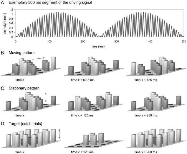
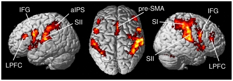
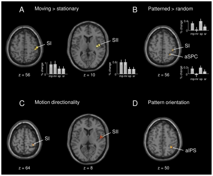
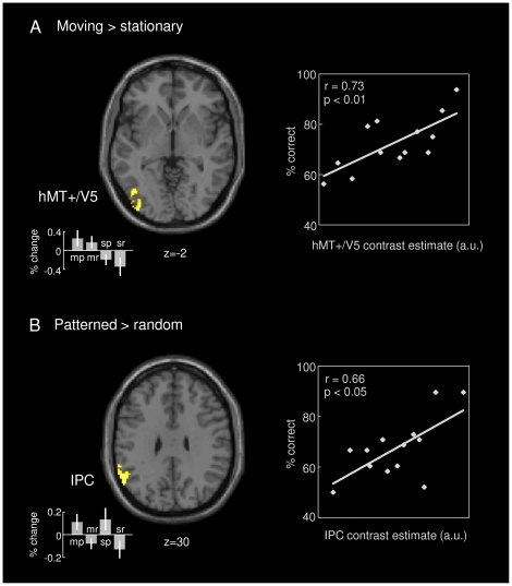
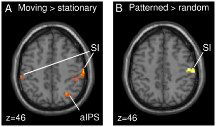
Processing of motion and pattern has been extensively studied in the visual domain, but much less in the somatosensory system. Here, we used ultra-high-field functional magnetic resonance imaging (fMRI) at 7 Tesla to investigate the neuronal correlates of tactile motion and pattern processing in humans under tightly controlled stimulation conditions. Different types of dynamic stimuli created the sensation of moving or stationary bar patterns during passive touch. Activity in somatosensory cortex was increased during both motion and pattern processing and modulated by motion directionality in primary and secondary somatosensory cortices (SI and SII) as well as by pattern orientation in the anterior intraparietal sulcus. Furthermore, tactile motion and pattern processing induced activity in the middle temporal cortex (hMT+/V5) and in the inferior parietal cortex (IPC), involving parts of the supramarginal und angular gyri. These responses covaried with subjects' individual perceptual performance, suggesting that hMT+/V5 and IPC contribute to conscious perception of specific tactile stimulus features. In addition, an analysis of effective connectivity using psychophysiological interactions (PPI) revealed increased functional coupling between SI and hMT+/V5 during motion processing, as well as between SI and IPC during pattern processing. This connectivity pattern provides evidence for the direct engagement of these specialized cortical areas in tactile processing during somesthesis.


Conflict of interest statement

Competing Interests: The authors have declared that no competing interests exist.

Figures

Figure 1
Figure 1. Illustration of tactile stimuli used.
A. The pins' driving signal was a 144 Hz sinusoidal carrier, which was amplitude-modulated by a rectified 2 Hz sine function. B. A directed propagation of the diagonals' sine phase across the display plane resulted in a percept of a bar pattern travelling smoothly across the fingertip. Both upward and downward diagonal orientations were used corresponding to orthogonal moving directions. For moving random stimuli, each of the four sine phases was assigned to four randomly chosen pins (not shown). C. Diagonals oscillating at opposite phases created the percept of a stationary bar pattern, which was periodically elevated and retracted. Again, both diagonal orientations were used. For stationary random stimuli, the driving signals were assigned to sets of randomly chosen pins (not shown). D. The target stimulus (to be detected on infrequently presented “catch” trials) was an oscillating square.
Figure 2
Figure 2. Overall neuronal network associated with tactile stimulation.
Contrasting tactile stimulation trials with null events revealed a distributed network involved in tactile information processing, including contralateral SI and bilateral SII, anterior intraparietal sulcus (aIPS), inferior frontal gyrus (IFG), lateral prefrontal cortex (LPFC), pre-supplementary motor area (pre-SMA), insular cortex, thalamus, and cerebellum. (Group-level analysis; pcluster<0.05, whole-brain FWE corr.).
Figure 3
Figure 3. Motion- and pattern-specific differences and differential effects for motion direction and pattern orientation.
A–B. Differential effects within the network associated with tactile stimulation (shown in Figure 2). Contrasting moving with stationary trials revealed an increased BOLD response in contralateral SI and SII (A). Contrasting patterned with random stimulus trials showed an increased BOLD response in anterior superior parietal cortex (aSPC) and SI (B). Effect sizes are plotted in terms of % signal change for all four stimulus types: moving patterned (mp), moving random (mr), stationary patterned (sp), and stationary random (sr). C–D. Differential effects for motion direction and pattern orientation. The regressor for moving pattern orientation revealed directionality effects for moving patterns in SI and SII (C). Differential effects for pattern orientation of stationary patterns were found in anterior intraparietal sulcus (aIPS; D). (Group-level analysis; p<0.005, uncorr.).
Figure 4
Figure 4. Areas involved in tactile motion and pattern processing.
A. Contrasting moving with stationary trials revealed an increased BOLD response in medial superior parietal cortex (not visible) and middle temporal cortex (hMT+/V5; on the left). Individual subjects' contrast estimates in hMT+/V5 correlated positively with their accuracy in identifying moving stimuli correctly (on the right). B. Contrasting patterned with random stimulus trials revealed an increased BOLD response in inferior parietal cortex (IPC; on the left). Individual subjects' contrast estimates in IPC correlated positively with their accuracy in identifying patterned stimuli correctly (on the right). A-B. Effect sizes are plotted in terms of % signal change for all four stimulus types: moving patterned (mp), moving random (mr), stationary patterned (sp), and stationary random (sr). (Group-level analysis; pcluster<0.05, whole-brain FWE corr.).
Figure 5
Figure 5. Psychophysiological interaction analyses using left hMT+/V5 and left IPC as seed regions.
A. The interaction term for moving vs. stationary trials revealed a significant increase in coupling between left hMT+/V5, bilateral SI, and right anterior intraparietal sulcus (aIPS) during motion processing. B. For patterned vs. random stimulus trials, the coupling between left IPC and right SI was significantly increased. (Group-level analysis; pcluster<0.05, whole-brain FWE corr.).

References

    1. Penfield W, Boldrey E. Somatic motor and sensory representation in the cerebral cortex of man as studied by electrical stimulation. Brain. 1937;60:389–443.
    1. Whitsel BL, Petrucelli LM, Werner G. Symmetry and connectivity in the map of the body surface in somatosensory area II of primates. J Neurophysiol. 1969;32:170–183. - PubMed
    1. Merzenich MM, Kaas JH, Sur M, Lin CS. Double representation of the body surface within cytoarchitectonic areas 3b and 1 in ‘SI’ in the owl monkey (Aotus trivirgatus). J Comp Neurol. 1978;181:41–73. - PubMed
    1. Kaas JH, Nelson RJ, Sur M, Lin CS, Merzenich MM. Multiple representations of the body within the primary somatosensory cortex of primates. Science. 1979;204:521–523. - PubMed
    1. Nelson RJ, Sur M, Felleman DJ, Kaas JH. Representations of the body surface in postcentral parietal cortex of Macaca fascicularis. J Comp Neurol. 1980;192:611–643. - PubMed

Publication types