. 2011;6(9):e24961.
doi: 10.1371/journal.pone.0024961. Epub 2011 Sep 16.

Differential effects of peptidoglycan recognition proteins on experimental atopic and contact dermatitis mediated by Treg and Th17 cells

Affiliations

Differential effects of peptidoglycan recognition proteins on experimental atopic and contact dermatitis mediated by Treg and Th17 cells

Shin Yong Park et al. PLoS One. 2011.

Abstract

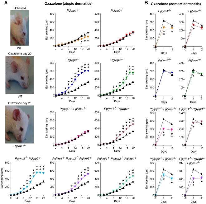
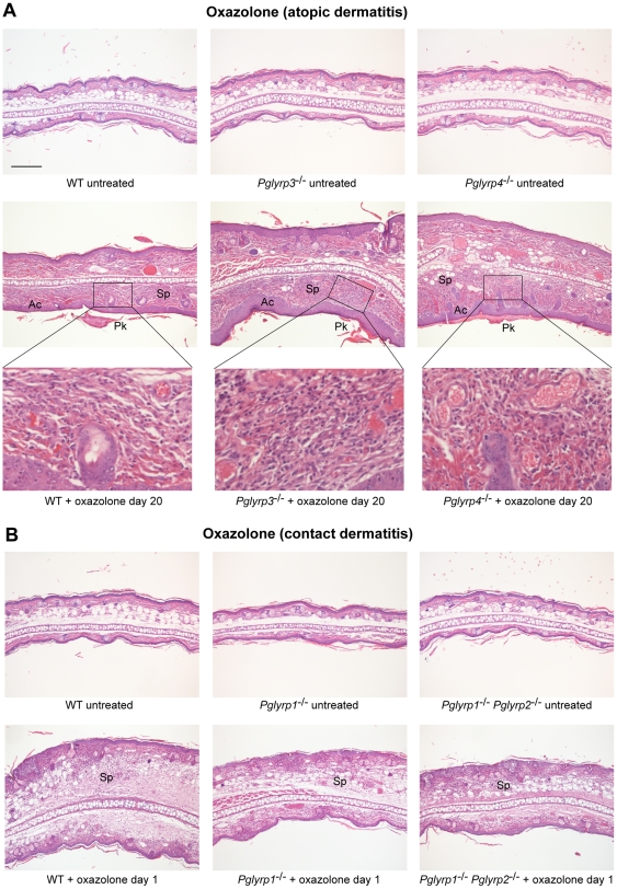
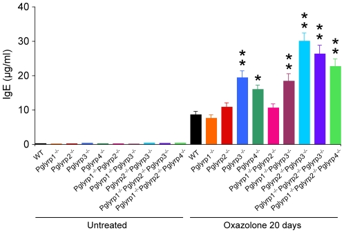
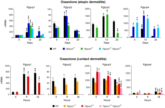
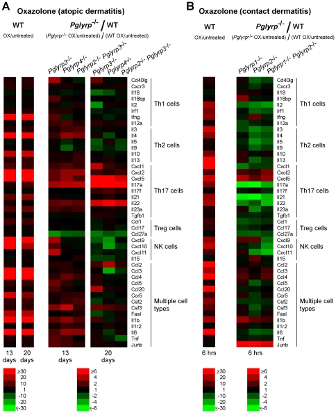
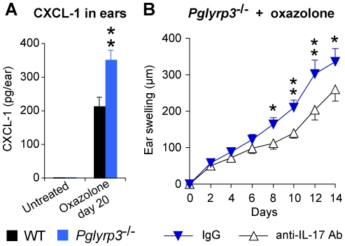
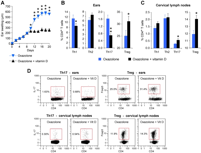
Skin protects the body from the environment and is an important component of the innate and adaptive immune systems. Atopic dermatitis and contact dermatitis are among the most frequent inflammatory skin diseases and are both determined by multigenic predisposition, environmental factors, and aberrant immune response. Peptidoglycan Recognition Proteins (Pglyrps) are expressed in the skin and we report here that they modulate sensitivity to experimentally-induced atopic dermatitis and contact dermatitis. Pglyrp3(-/-) and Pglyrp4(-/-) mice (but not Pglyrp2(-/-) mice) develop more severe oxazolone-induced atopic dermatitis than wild type (WT) mice. The common mechanism underlying this increased sensitivity of Pglyrp3(-/-) and Pglyrp4(-/-) mice to atopic dermatitis is reduced recruitment of Treg cells to the skin and enhanced production and activation Th17 cells in Pglyrp3(-/-) and Pglyrp4(-/-) mice, which results in more severe inflammation and keratinocyte proliferation. This mechanism is supported by decreased inflammation in Pglyrp3(-/-) mice following in vivo induction of Treg cells by vitamin D or after neutralization of IL-17. By contrast, Pglyrp1(-/-) mice develop less severe oxazolone-induced atopic dermatitis and also oxazolone-induced contact dermatitis than WT mice. Thus, Pglyrp3 and Pglyrp4 limit over-activation of Th17 cells by promoting accumulation of Treg cells at the site of chronic inflammation, which protects the skin from exaggerated inflammatory response to cell activators and allergens, whereas Pglyrp1 has an opposite pro-inflammatory effect in the skin.


Conflict of interest statement

Competing Interests: The authors have declared that no competing interests exist.

Figures

Figure 1
Figure 1. Pglyrp3 −/− and Pglyrp4 −/− mice have enhanced response in the oxazolone model of atopic dermatitis and Pglyrp1 −/− and other Pglyrp −/− mice have reduced response in the oxazolone model of contact dermatitis.
(A) Atopic dermatitis model: sensitization followed by 10 applications of oxazolone to the ears every other day induces mild inflammation (left) and mild ear swelling (right) in WT mice (black triangles) and severe inflammation with increased redness, scaling, and extensive ear swelling in Pglyrp3 −/− and Pglyrp4 −/− mice (color triangles). (B) Contact dermatitis model: sensitization followed by a single application of oxazolone to the ears induces strong ear swelling in WT mice (black triangles) and reduced ear swelling in Pglyrp1 −/−, Pglyrp2 −/−, and Pglyrp4 −/− mice (color triangles). Means ± SEM (SEM were often smaller than the symbols in this and other figures); N = 9–17 mice/group; significance of differences between Pglyrp −/− and WT mice: *, P<0.02; **, P<0.0001.
Figure 2
Figure 2. Ear histology in WT and Pglyrp-deficient mice in atopic dermatitis and contact dermatitis models of skin inflammation.
(A) Oxazolone model of atopic dermatitis: sensitization and 10 applications of oxazolone to the ears every other day induced acanthosis (Ac), parakeratosis (Pk), marked thickening of the sub-epidermal layer with spongiosis (Sp) and dense cellular infiltrates of primarily mononuclear and some polymorphonuclear cells (high magnification insets), that were all highly prominent in Pglyrp3 −/− mice and Pglyrp4 −/− mice and much less severe in WT mice. (B) Oxazolone model of contact dermatitis: sensitization and a single application of oxazolone to the ears induced strong inflammatory response in WT mice with marked spongiosis of the sub-epidermal layer (Sp) and cellular infiltrates of epidermal and sub-epidermal layers, composed of mononuclear and polymorphonuclear cells; Pglyrp1 −/− and Pglyrp1 −/− Pglyrp2 −/− mice still had cellular infiltrates, but had substantially reduced swelling, compared to WT mice, mostly due to reduced edema. H&E stained cross-sections; bar = 200 µm for all panels, except high magnification insets (the magnified areas are shown by rectangles).
Figure 3
Figure 3. Pglyrp3 −/− and Pglyrp4 −/− mice have higher serum IgE levels than WT mice in the oxazolone model of atopic dermatitis.
Sensitization followed by 10 applications of oxazolone to the ears every other day induced higher level of serum IgE in Pglyrp3 −/− and Pglyrp4 −/− mice than in WT, Pglyrp1 −/− or Pglyrp2 −/− mice; means ± SEM of 8–20 mice/group; *, P<0.002; **, P<0.0001, Pglyrp −/− versus WT.
Figure 4
Figure 4. Pglyrp1, Pglyrp2, Pglyrp3, and Pglyrp4 expression is increased in the affected skin in the atopic dermatitis and contact dermatitis models of inflammation.
The amounts of each PGRP mRNA in WT mice and in the indicated Pglyrp-deficient mice sensitized and treated with oxazolone every other day (atopic dermatitis model) or sensitized and treated with oxazolone at 0 hrs only (contact dermatitis model) were measured by qRT-PCR. The results are means of 3–4 mice ± SEM; *, P<0.04; **, P<0.001, treated versus untreated; ♦, P<0.04; ♦♦, P<0.001, Pglyrp −/− versus WT.
Figure 5
Figure 5. Pglyrp3 and Pglyrp4 are expressed primarily in the differentiated epidermal cells in both untreated and oxazolone-treated skin in the atopic dermatitis model.
Transverse sections of the ears from either untreated or oxazolone-treated mice (sensitization followed by 10 applications of oxazolone to the ears every other day) were stained by an immunoperoxidase method with either anti-Pglyrp3 or anti-Pglyrp4 antibodies or IgG (negative control); bar = 50 µm.
Figure 6
Figure 6. Pglyrp3 −/− and Pglyrp4 −/− mice have increased Th17 cells in the affected skin in the atopic dermatitis model, whereas Pglyrp1 −/− and Pglyrp2 −/− mice have most cell types decreased (except B cells and Treg cells) in the contact dermatitis model.
Expression of a panel of marker genes characteristic of various inflammatory cell types in the ears of mice after (A) sensitization and 10 applications of oxazolone to the ears every other day, or (B) after sensitization and 6 hrs after a single application of oxazolone to the ears, measured by qRT-PCR is shown. For WT mice (left panels in A and B), the ratio of the amount of mRNA in oxazolone-treated to untreated mice for each gene (fold induction by oxazolone) is shown; for Pglyrp −/− mice (right panels in A and B), the results are the ratios of fold induction of each gene by oxazolone in Pglyrp −/− mice to fold induction of each gene by oxazolone in WT mice (which represents the fold difference in the response to oxazolone in Pglyrp −/− versus WT mice). The results are means of 3 arrays from 4–5 mice/group in heat map format. The means ± SEM bar graphs for these results are shown in Figures S1 and S2.
Figure 7
Figure 7. Th17 gene expression profile is preferentially induced in the atopic dermatitis model in Pglyrp3 −/− and Pglyrp4 −/− mice, whereas expression of most immune genes is reduced in the contact dermatitis model in Pglyrp −/− mice.
Expression of a panel of cytokines, chemokines, and other marker genes characteristic of Th1, Th2, Th17, Treg, NK, and other cell types in the ears of mice was measured by qRT-PCR. (A) After sensitization and 10 applications of oxazolone to the ears every other day the expression of several Th17 marker genes was higher in Pglyrp3 −/− and Pglyrp4 −/− mice than in WT mice. (B) After sensitization and a single application of oxazolone to the ears the expression of most of immune marker genes was lower in Pglyrp −/− mice than in WT mice. For WT mice (left panels in A and B), the ratio of the amount of mRNA in oxazolone-treated to untreated mice for each gene (fold induction by oxazolone) is shown; for Pglyrp −/− mice (right panels in A and B), the results are the ratios of fold induction of each gene by oxazolone in Pglyrp −/− mice to fold induction of each gene by oxazolone in WT mice (which represents the fold difference in the response to oxazolone in Pglyrp −/− versus WT mice). The results are means of 3 arrays from 4–5 mice/group in heat map format. The means ± SEM bar graphs for these results are shown in Figures S3, S4, and S5.
Figure 8
Figure 8. Pglyrp3 −/− mice have high numbers Th17 and low numbers of Treg cells in the affected skin in the oxazolone model of atopic dermatitis.
(A) Numbers of CD4+ cells and Th17 cells in the ears or (B–D) percentages of Th1, Th2, Th17, and Treg cells in the ears, cervical lymph nodes, and spleen in sensitized WT and Pglyrp3 −/− mice on days 13 or 20 of ear treatment with oxazolone, measured by flow cytometry; means ± SEM of 5–9 mice/group (*, P<0.05; **, P<0.005; Pglyrp3 −/− versus WT) or representative dot plots are shown. (E) Expression of receptors for chemokines that attract Treg cells in cervical lymph nodes of sensitized WT and Pglyrp3 −/− mice on day 20 of ear treatment with oxazolone measured by qRT-PCR; amounts of mRNA are shown as means ± SEM of 3 arrays from 5 mice/group.
Figure 9
Figure 9. IL-17 is required for enhanced response to oxazolone in Pglyrp3 −/− mice.
(A) The level of IL-17-induced chemokine, CXCL-1, is higher in the ears of Pglyrp3 −/− mice than WT mice after sensitization and application of oxazolone for 20 days. (B) Ear swelling in Pglyrp3 −/− mice sensitized and treated 7 times with oxazolone every other day and also treated with neutralizing anti-IL-17 mAbs is lower than in Pglyrp3 −/−mice similarly treated with oxazolone and isotype control IgG. Means ± SEM; N = 6 mice/group; significance of differences between Pglyrp3 −/− and WT mice (A) or IgG control and anti-IL-17 mAbs-treated mice (B): *, P<0.05; **, P<0.005.
Figure 10
Figure 10. Induction of Treg cells by vitamin D reduces the inflammatory response to oxazolone and decreases the numbers of Th17 cells in the skin of Pglyrp3 −/− mice.
(A) Vitamin D applied to the skin of Pglyrp3 −/− mice together with oxazolone reduces ear swelling compared to Pglyrp3 −/− mice similarly treated with oxazolone alone. (B–D) Vitamin D applied to the skin of Pglyrp3 −/− mice together with oxazolone increases the percentages of Treg cells in the ears and cervical lymph nodes and reduces the percentages of Th17 cells in the ears compared to the application of oxazolone alone, measured on day 20 by flow cytometry. Means ± SEM of 8 mice/group (A–C; *, P<0.05; **, P<0.005; oxazolone versus oxazolone + vitamin D) or representative dot plots (D) are shown.

Similar articles

Cited by

References

    1. De Benedetto A, Agnihothri R, McGirt LY, Bankova LG, Beck LA. Atopic dermatitis: a disease caused by innate immune defects? J Invest Dermatol. 2009;129:14–30. - PubMed
    1. Fonacier LS, Dreskin SC, Leung DY. Allergic skin diseases. J Allergy Clin Immunol. 2010;125(Suppl 2):S138–149. - PubMed
    1. Jin H, He R, Oyoshi M, Geha RS. Animal models of atopic dermatitis. J Invest Dermatol. 2009;129:31–40. - PMC - PubMed
    1. Wang B, Feliciani C, Freed I, Cai Q, Sauder DN. Insights into molecular mechanisms of contact hypersensitivity gained from gene knockout studies. J Leukoc Biol. 2001;70:185–191. - PubMed
    1. Martin SF, Jakob T. From innate to adaptive immune responses in contact hypersensitivity. Curr Opin Allergy Clin Immunol. 2008;8:289–293. - PubMed

Publication types

MeSH terms