

Administration of simvastatin after kainic acid-induced status epilepticus restrains chronic temporal lobe epilepsy
- PMID: 21949812
- PMCID: PMC3176286
- DOI: 10.1371/journal.pone.0024966
Administration of simvastatin after kainic acid-induced status epilepticus restrains chronic temporal lobe epilepsy
Abstract
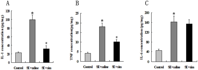
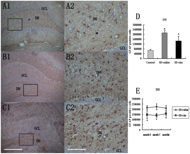
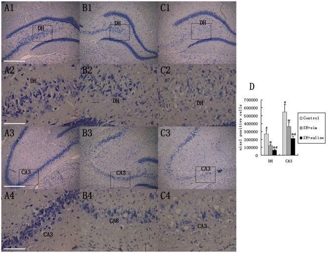
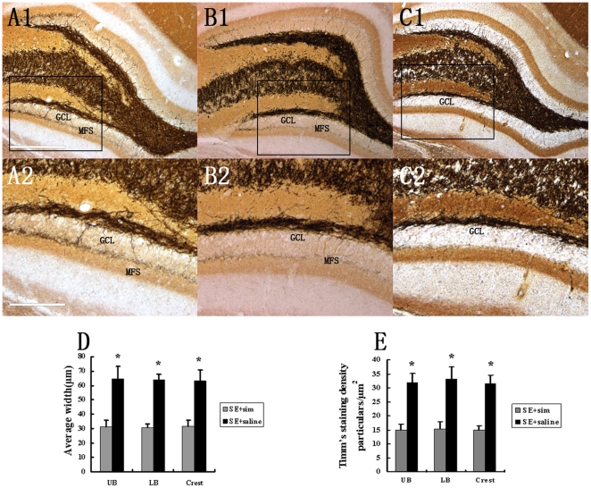
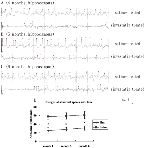
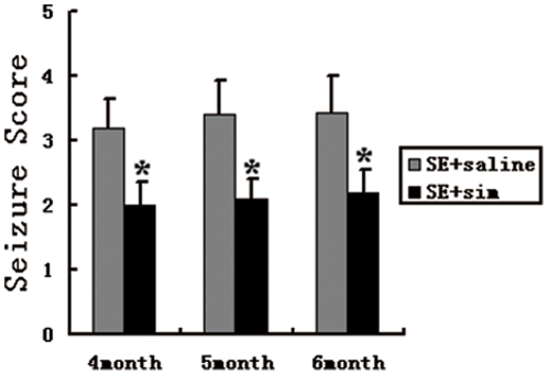
In this study, we examined the effect of chronic administration of simvastatin immediately after status epilepticus (SE) on rat brain with temporal lobe epilepsy (TLE). First, we evaluated cytokines expression at 3 days post KA-lesion in hippocampus and found that simvastatin-treatment suppressed lesion-induced expression of interleukin (IL)-1β and tumor necrosis factor-α (TNF-α). Further, we quantified reactive astrocytosis using glial fibrillary acidic protein (GFAP) staining and neuron loss using Nissl staining in hippocampus at 4-6 months after KA-lesion. We found that simvastatin suppressed reactive astrocytosis demonstrated by a significant decrease in GFAP-positive cells, and attenuated loss of pyramidal neurons in CA3 and interneurons in dentate hilar (DH). We next assessed aberrant mossy fiber sprouting (MFS) that is known to contribute to recurrence of spontaneous seizure in epileptic brain. In contrast to the robust MFS observed in saline-treated animals, the extent of MFS was restrained by simvastatin in epileptic rats. Attenuated MFS was related to decreased neuronal loss in CA3 and DH, which is possibly a mechanism underlying decreased hippocampal susceptibility in animal treated with simvastatin. Electronic encephalography (EEG) was recorded during 4 to 6 months after KA-lesion. The frequency of abnormal spikes in rats with simvastatin-treatment decreased significantly compared to the saline group. In summary, simvastatin treatment suppressed cytokines expression and reactive astrocytosis and decreased the frequency of discharges of epileptic brain, which might be due to the inhibition of MFS in DH. Our study suggests that simvastatin administration might be a possible intervention and promising strategy for preventing SE exacerbating to chronic epilepsy.
Conflict of interest statement
Figures
Similar articles
-
Anoxia during kainate status epilepticus shortens behavioral convulsions but generates hippocampal neuron loss and supragranular mossy fiber sprouting.Epilepsy Res. 1998 Apr;30(2):133-51. doi: 10.1016/s0920-1211(97)00103-4. Epilepsy Res. 1998. PMID: 9600545
-
Temporal profile of clinical signs and histopathologic changes in an F-344 rat model of kainic acid-induced mesial temporal lobe epilepsy.Toxicol Pathol. 2008 Dec;36(7):932-43. doi: 10.1177/0192623308326093. Toxicol Pathol. 2008. PMID: 19126789
-
Hippocampal neurodegeneration, spontaneous seizures, and mossy fiber sprouting in the F344 rat model of temporal lobe epilepsy.J Neurosci Res. 2006 May 1;83(6):1088-105. doi: 10.1002/jnr.20802. J Neurosci Res. 2006. PMID: 16493685
-
The kainic acid model of temporal lobe epilepsy.Neurosci Biobehav Rev. 2013 Dec;37(10 Pt 2):2887-99. doi: 10.1016/j.neubiorev.2013.10.011. Epub 2013 Oct 30. Neurosci Biobehav Rev. 2013. PMID: 24184743 Free PMC article. Review.
-
Progress in neuroprotective strategies for preventing epilepsy.Prog Neurobiol. 2008 Apr;84(4):363-404. doi: 10.1016/j.pneurobio.2007.10.010. Epub 2007 Dec 8. Prog Neurobiol. 2008. PMID: 18207302 Free PMC article. Review.
Cited by
-
Lovastatin, not Simvastatin, Corrects Core Phenotypes in the Fragile X Mouse Model.eNeuro. 2019 Jun 12;6(3):ENEURO.0097-19.2019. doi: 10.1523/ENEURO.0097-19.2019. Print 2019 May/Jun. eNeuro. 2019. PMID: 31147392 Free PMC article.
-
Novel frontiers in epilepsy treatments: preventing epileptogenesis by targeting inflammation.Expert Rev Neurother. 2013 Jun;13(6):615-25. doi: 10.1586/ern.13.54. Expert Rev Neurother. 2013. PMID: 23738999 Free PMC article. Review.
-
Lovastatin modulates glycogen synthase kinase-3β pathway and inhibits mossy fiber sprouting after pilocarpine-induced status epilepticus.PLoS One. 2012;7(6):e38789. doi: 10.1371/journal.pone.0038789. Epub 2012 Jun 26. PLoS One. 2012. PMID: 22761705 Free PMC article.
-
The potential of antiseizure drugs and agents that act on novel molecular targets as antiepileptogenic treatments.Neurotherapeutics. 2014 Apr;11(2):385-400. doi: 10.1007/s13311-014-0266-1. Neurotherapeutics. 2014. PMID: 24671870 Free PMC article. Review.
-
Myoloid-related protein 8, an endogenous ligand of Toll-like receptor 4, is involved in epileptogenesis of mesial temporal lobe epilepsy via activation of the nuclear factor-κB pathway in astrocytes.Mol Neurobiol. 2014 Feb;49(1):337-51. doi: 10.1007/s12035-013-8522-7. Epub 2013 Aug 28. Mol Neurobiol. 2014. PMID: 23982744
References
-
- Strine TW, Kobau R, Chapman DP, Thurman DJ, Price P, et al. Psychological distress, comorbidities, and health behaviors among U. S. adults with seizures: results from the 2002 National Health Interview Survey. Epilepsia. 2005;46:1133–1139. - PubMed
-
- Litt B, Esteller R, Echauz J, D'Alessandro M, Shor R, et al. Epileptic seizures may begin hours in advance of clinical onset: a report of five patients. Neuron. 2001;30:51–64. - PubMed
-
- Manford M, Hart YM, Sander JW, Shorvon SD. National General Practice Study of Epilepsy: partial seizure patterns in a general population. Neurology. 1992;42:1911–1917. - PubMed
-
- Lieb JP, Babb TL, Engel J., Jr Quantitative comparison of cell loss and thiopental-induced EEG changes in human epileptic hippocampus. Epilepsia. 1989;30:147–156. - PubMed
-
- French JA, Williamson PD, Thadani VM, Darcey TM, Mattson RH, et al. Characteristics of medial temporal lobe epilepsy: Results of history and physical examination. Ann Neurol. 1993;34:774–780. - PubMed
Publication types
MeSH terms
Substances
LinkOut - more resources
Full Text Sources
Other Literature Sources
Medical
Miscellaneous
