. 2011;6(9):e25108.
doi: 10.1371/journal.pone.0025108. Epub 2011 Sep 20.

Type III Nrg1 back signaling enhances functional TRPV1 along sensory axons contributing to basal and inflammatory thermal pain sensation

Affiliations

Type III Nrg1 back signaling enhances functional TRPV1 along sensory axons contributing to basal and inflammatory thermal pain sensation

Sarah E Canetta et al. PLoS One. 2011.

Abstract

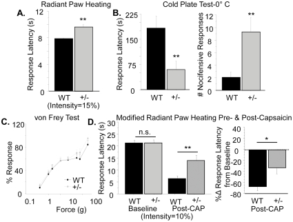
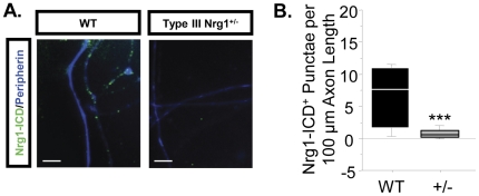
Type III Nrg1, a member of the Nrg1 family of signaling proteins, is expressed in sensory neurons, where it can signal in a bi-directional manner via interactions with the ErbB family of receptor tyrosine kinases (ErbB RTKs). Type III Nrg1 signaling as a receptor (Type III Nrg1 back signaling) can acutely activate phosphatidylinositol-3-kinase (PtdIns3K) signaling, as well as regulate levels of α7* nicotinic acetylcholine receptors, along sensory axons. Transient receptor potential vanilloid 1 (TRPV1) is a cation-permeable ion channel found in primary sensory neurons that is necessary for the detection of thermal pain and for the development of thermal hypersensitivity to pain under inflammatory conditions. Cell surface expression of TRPV1 can be enhanced by activation of PtdIns3K, making it a potential target for regulation by Type III Nrg1. We now show that Type III Nrg1 signaling in sensory neurons affects functional axonal TRPV1 in a PtdIns3K-dependent manner. Furthermore, mice heterozygous for Type III Nrg1 have specific deficits in their ability to respond to noxious thermal stimuli and to develop capsaicin-induced thermal hypersensitivity to pain. Cumulatively, these results implicate Type III Nrg1 as a novel regulator of TRPV1 and a molecular mediator of nociceptive function.


Conflict of interest statement

Competing Interests: The authors have declared that no competing interests exist.

Figures

Figure 1
Figure 1. Type III Nrg1 expression in adult nociceptive sensory neuron soma and peripheral nerve terminals in vivo.
(A) Type III Nrg1 (b,e,h) is found in nociceptive TrkA+ (a–c) and IB4+ (d–f), as well as TRPV1+ (g–i) sensory neuron soma in lumbar dorsal root ganglia. (B) Type III Nrg1 (b′,e′) is found in nociceptive peripheral nerve terminals innervating the plantar hindpaw skin as marked with an antibody against CGRP (a′) as well as TRPV1 (d′). White arrows indicate examples of soma or nerve terminals that are expressing both Type III Nrg1 and the particular sensory marker. All scale bars equal 10 µm.
Figure 2
Figure 2. Type III Nrg1+/− animals have specific behavioral deficits in responding to noxious thermal stimuli.
(A) Response in radiant paw heating assay. Latency to respond to radiant heat (set at 15% of maximum intensity) applied to the hindpaw was evaluated for WT and Type III Nrg1+/− siblings. Response times from both paws were averaged by animal and compared by genotype. Type III Nrg1+/− mice showed a significantly increased latency to respond relative to their WT littermates (WT, n = 16 animals; Type III Nrg1+/−, n = 14 animals; **p<0.01). (B) Response to noxious cold assessed with the cold plate test. Latency to respond to a 0°C stimulus was measured for WT and Type III Nrg1+/− mice and compared by genotype. Type III Nrg1+/− mice respond significantly faster than their WT littermates (WT, n = 16 animals; Type III Nrg1+/−, n = 14 animals; **p<0.01). The total number of nocifensive responses that each animal made in the 5 minute period they remained on the 0°C plate was measured and compared by genotype. Type III Nrg1+/− mice made significantly more nocifensive responses than their WT littermates (**p<0.01). (C) Response in the von Frey assay of mechanosensation. Percent response for the various forces was evaluated for WT and Type III Nrg1+/− mice. Data were compared by genotype for each test force. There were no significant differences between genotypes (WT, n = 15 animals; Type III Nrg1+/−, n = 14 animals). (D) Response in a modified version of the radiant paw heating assay pre- and post-capsaicin application. Latency to respond to radiant heat (set at 10% of maximum intensity) applied to the hindpaw was evaluated for WT and Type III Nrg1+/− littermates before and after application of 15 µl of 0.075% capsaicin cream to the right hindpaw. Response latencies following capsaicin application were significantly less depressed in Type III Nrg1+/− animals relative to WTs (WT, n = 16 animals; Type III Nrg1+/−, n = 13 animals **p<0.01). Similarly, the percent change (%Δ) in response latency for Type III Nrg1+/− animals was significantly blunted relative to WTs (*p<0.05). All genotype comparisons were made using a Student's t-test. All graphs show the mean±SEM.
Figure 3
Figure 3. Adult WT and Type III Nrg1+/− animals have equivalent numbers of sensory neurons and sensory cutaneous projections.
(A) Representative sections through L4/L5 DRG from adult WT and Type III Nrg1+/− mice were stained with a pan-sensory marker. Total cells staining positive for this marker tallied from at least 5 sections evenly spaced throughout the DRG were counted and averaged for each animal. Genotype averages were compared with a Student's t-test (n = 3 animals per genotype). There was no statistically significant difference between genotypes. (B) Galabrous hindpaw skin samples from WT and Type III Nrg1+/− mice were assessed for total TrkA, Ret and TRPV1 protein using immunoblot. (C) The intensity of the TrkA, Ret and TRPV1 bands were quantified and normalized to GAPDH to control for equal protein loading. The values were expressed as a fold change from the average WT value and genotype averages were compared with a Student's t-test (for TrkA and Ret: WT n = 13 paws from 10 animals, Type III Nrg1+/− n = 13 paws from 9 animals; for TRPV1: WT n = 9 paws from 5 animals, Type III Nrg1+/− n = 8 paws from 5 animals). There was no statistically significant difference between the genotypes.
Figure 4
Figure 4. Sensory axons, but not soma, from Type III Nrg1+/− mice show reduced capsaicin responsiveness compared to axons from WT mice.
(A) Representative traces of intracellular calcium along sensory axons in response to 1 µM capsaicin or 56 mM KCl. The change in intracellular calcium from baseline over time ([(F−F0)/F0]*100) is shown for WT (left) and Type III Nrg1+/− (right) axons. Hatched diagonal lines indicate where the time course was non-continuous. (B) Quantification of the maximum change in intracellular calcium in response to application of 1 µM capsaicin or 56 mM KCl by genotype. Averages of 5 animals per genotype were compared using a Student's t-test. Type III Nrg1+/− axons showed a significantly decreased response to capsaicin (p<0.05), but not to KCl, relative to WTs. Graph shows mean±SEM. (C) Type III Nrg1+/− sensory soma show normal response to capsaicin. Quantification of maximal change in fluorescence from baseline ([(F−F0)/F0]*100) in WT or Type III Nrg1+/− sensory neuron soma in response to 1 µM capsaicin or 56 mM KCl. Average responses from 4 WT and 4 Type III Nrg1+/− animals to application of capsaicin or KCl were compared by genotype using a Student's t-test. There was no statistically significant difference between genotypes. Graphs show mean±SEM. (D) Type III Nrg1 (green) and TRPV1 (red) are co-expressed along P21 WT cultured sensory neuron axons identified with a pan-axonal (PA) marker (blue). White arrows indicate examples where Type III Nrg1 and TRPV1 are in close proximity. Scale bar equals 10 µm. (E) P21 WT and Type III Nrg1+/− sensory neuron cultures have equivalent levels of total TRPV1 protein. Total TRPV1 protein measurement by immunoblot. The 95 kD TRPV1 band and the 35 kD GAPDH band are shown from a representative experiment comparing protein from P21 WT and Type III Nrg1+/− cultures. Quantification of fold change in intensity of TRPV1∶GAPDH normalized to WT average. There was no statistically significant change in the ratio of TRPV1 to GAPDH between genotypes (WT, Type III Nrg1+/−, n = 3 animals). Genotype comparisons were made using a Student's t-test. Graph shows mean±SEM.
Figure 5
Figure 5. Stimulation of Type III Nrg1 signaling enhances response to capsaicin in WT sensory axons.
(A) Representative traces of intracellular calcium along WT sensory axons in response to repeated applications of 1 µM capsaicin followed by application of 56 mM KCl. The maximum percent change in intracellular calcium ([(F−F0)/F0]*100) in response to capsaicin decreased between the 4th and 5th capsaicin applications under control conditions (left), but increased when Type III Nrg1 signaling was stimulated by sErbB4-ECD application during that interval (right). (B) Quantification of percent change in maximum response to capsaicin between the 4th and the 5th capsaicin application ([(F5−F4)/F4]*100) by treatment. WT sensory axons showed a significantly enhanced response to capsaicin when Type III Nrg1 signaling was stimulated with sErbB4-ECD (WT CON, n = 10 animals; WT B4, n = 7 animals; Student's t-test, ***p<0.001). Graph shows mean±SEM.
Figure 6
Figure 6. PtdIns3K activation is required for Type III Nrg1-induced enhancement of capsaicin responsiveness.
(A) 15-minute stimulation of Type III Nrg1 signaling with sErbB4-ECD significantly increased the ratio of pAKT-AKT in whole cell lysates from P21 WT sensory neuron cultures (a: CON, n = 3; B4, n = 4; Student's t-test, *p<0.05). This increase was blocked by pre-incubation with 20 nM wortmannin (WM), an inhibitor of PtdIns3K activity. (b) The same stimulation of Type III Nrg1 with sErbB4-ECD did not activate the MAPK pathway as illustrated by the lack of effect on the ratio of pERK-ERK. (B) Stimulation of Type III Nrg1 signaling with sErbB4-ECD significantly increased average fluorescence intensity (AFI) levels for pAKT staining along P21 WT cultured sensory axons (15 minutes; CON, n = 3; B4, n = 3; Student's t-test, *p<0.05). Scale bars equal 10 µm. (C) Blocking PtdIns3K signaling with 50 µM LY294002 or 20 nM wortmannin blocked the Type III Nrg1-induced enhancement of functional TRPV1 (LY CON, n = 3 animals; LY B4, n = 2; WM CON, n = 10; WM B4, n = 7). Comparisons between CON and all treatment groups were made using an ANOVA with a Holm-Sidak post-hoc test for multiple comparisons. All graphs show mean±SEM.
Figure 7
Figure 7. Pre-treatment with sErbB4-ECD enhances response to a second application of capsaicin along sensory axons.
(A) Sensory neuron cultures from P21 WT mice were incubated with sErbB4-ECD or control media for 12 minutes before two 1 µM pulses of capsaicin were applied (spaced 4 minutes apart), followed by application of 56 mM KCl. Within axons that responded to both capsaicin and KCl, the percent change in fluorescence from baseline ([(F−F0)/F0]*100) in response to both applications of capsaicin as well as KCl was calculated. Axonal responses were averaged by animal and normalized to the WT CON average. (B–C) Quantification of the maximum response to the first (B) or second (C) applications of 1 µM capsaicin were compared by treatment. Pre-treatment with sErbB4-ECD significantly increased the maximum response to the second pulse of capsaicin (WT CON, n = 8 animals; WT B4, n = 9 animals; *p<0.05). There was a trend towards an increase in response to the first pulse of capsaicin following sErbB4-ECD pre-treatment (p = 0.17). (D) Quantification of the maximum response to KCl by treatment. sErbB4-ECD pre-treatment had no effect on the maximum response to KCl (p = 0.38). (E) The percent change between the maximum response to the first and second capsaicin applications was calculated and axonal values were averaged by animal. Quantification of this percent change in response by treatment; Cultures pre-treated with sErbB4-ECD showed significantly less of a decrease in responsiveness to capsaicin between the first and second capsaicin applications (*p<0.05). All treatment comparisons were made using a Student's t-test. Graphs show mean±SEM.
Figure 8
Figure 8. Blockade of ErbB signaling does not enhance response to capsaicin in WT sensory axons.
(A) Incubation of P21 WT sensory neuron cultures with soluble Nrg1 (sNrg1) stimulates ErbB signaling and activates ERK (measured as a ratio of pERK to GAPDH). This sNrg1-induced activation of ERK can be blocked by incubation with 1 µM PD158780, an ErbB kinase inhibitor (n = 3 animals per condition; *p<0.05). (B–D) Blockade of ErbB signaling does not enhance response to application of capsaicin or KCl. Sensory neuron cultures from P21 WT mice were incubated with 1 µM PD158780 or control media for 12 minutes before two 1 µM pulses of capsaicin were applied (spaced 4 minutes apart), followed by application of 56 mM KCl. Within axons that responded to both capsaicin and KCl, the percent change in fluorescence from baseline ([(F−F0)/F0]*100) in response to both applications of capsaicin as well as KCl was calculated. Axonal responses were averaged by animal and normalized to the WT CON average. Quantification of the maximum response to the first (B) or second (C) applications of 1 µM capsaicin or KCl (D) were compared by treatment. Blockade of ErbB signaling with 1 µM PD158780 did not affect response to the first or second application of capsaicin, or to KCl (WT CON, n = 7 animals; ErbB Inh, n = 7 animals).
Figure 9
Figure 9. Acute stimulation of Type III Nrg1 signaling in Type III Nrg1+/− sensory axons does not rescue functional TRPV1 deficits.
(A) Representative traces of intracellular calcium along Type III Nrg1+/− sensory axons in response to repeated applications of 1 µM capsaicin followed by application of 56 mM KCl. The maximum percent change in intracellular calcium ([(F−F0)/F0]*100) in response to capsaicin decreased between the 4th and 5th capsaicin application in Type III Nrg1+/− axons under control conditions (left) and did not increase when Type III Nrg1 signaling was stimulated by sErbB4-ECD application during that interval (right). (B) Quantification of percent change in maximum response to capsaicin between the 4th and the 5th capsaicin application ([(F5−F4)/F4]*100) by treatment. Results from WT sensory axons are included for comparison (WT CON, n = 10; WT B4, n = 7; ***p<0.001). Type III Nrg1+/− axons did not show a statistically significantly enhanced response to capsaicin when Type III Nrg1 signaling was stimulated (Type III Nrg1+/− CON, n = 6 animals; Type III Nrg1+/− B4, n = 8). All comparisons between genotypes and treatments were made using an ANOVA with a Holm-Sidak post-hoc test for multiple comparisons. Graph shows mean±SEM.
Figure 10
Figure 10. sErbB4-ECD stimulation of Type III Nrg1+/− sensory neuron cultures fails to activate PtdIns3K along sensory axons.
(A) Representative images of pAKT staining along WT and Type III Nrg1+/− sensory axons stimulated for 15 minutes with either control or sErbB4-ECD media. All scale bars equal 10 µm. (B) Quantification of pAKT average fluorescence intensity (AFI) along WT and Type III Nrg1+/− sensory axons under various treatment conditions. sErbB4-ECD stimulation significantly increased AFI levels for pAKT staining along P21 WT sensory axons (WT CON, n = 3 animals; WT B4, n = 3; *p<0.05) but not along sensory axons from Type III Nrg1+/− cultures (Type III Nrg1+/− CON, n = 3 animals; Type III Nrg1+/− B4, n = 3; p = 0.9). All comparisons between treatments and genotypes were made using an ANOVA with a Fischer's PLSD post-hoc analysis. Graph shows mean±SEM.
Figure 11
Figure 11. Type III Nrg1+/− sensory axons have reduced numbers of Type III Nrg1+ punctae relative to WT sensory axons.
(A) Conventional microscopic images of Type III Nrg1+ punctae (green) found along peripherin+ axons (blue) from WT and Type III Nrg1+/− sensory cultures. Scale bars equal 10 µm. (B) Quantification of the average number of punctae per 100 µm axon length from 16 WT and 15 Type III Nrg1+/− images. Comparison by genotype illustrates that Type III Nrg1+/− sensory axons have significantly fewer Type III Nrg1+ punctae than WT sensory axons (Mann-Whitney Rank Sum test, *p<0.05).
Figure 12
Figure 12. Type III Nrg1+/− sensory axons have reduced numbers of Nrg1+ punctae that contain a signaling-competent intracellular domain (Nrg1-ICD) relative to WT sensory axons.
(A) Conventional microscopic images of Nrg1-ICD+ punctae (green) found along peripherin+ axons (blue) from WT and Type III Nrg1+/− sensory cultures. Scale bars equal 10 µm. (B) Quantification of the average number of punctae per 100 µm axon length from 14 WT and 15 Type III Nrg1+/− images. Comparison by genotype illustrates that Type III Nrg1+/− sensory axons have significantly fewer Nrg1-ICD+ punctae than WT sensory axons (Mann-Whitney Rank Sum test, ***p<0.001).

Similar articles

Cited by

References

    1. Bao J, Wolpowitz D, Role LW, Talmage DA. Back signaling by the Nrg-1 intracellular domain. J Cell Biol. 2003;161:1133–1141. - PMC - PubMed
    1. Hancock ML, Canetta SE, Role LW, Talmage DA. Presynaptic type III neuregulin1-ErbB signaling targets {alpha}7 nicotinic acetylcholine receptors to axons. J Cell Biol. 2008;181:511–521. - PMC - PubMed
    1. Caterina MJ, Leffler A, Malmberg AB, Martin WJ, Trafton J, et al. Impaired nociception and pain sensation in mice lacking the capsaicin receptor. Science. 2000;288:306–313. - PubMed
    1. Bonnington JK, McNaughton PA. Signalling pathways involved in the sensitisation of mouse nociceptive neurones by nerve growth factor. J Physiol. 2003;551:433–446. - PMC - PubMed
    1. Stein AT, Ufret-Vincenty CA, Hua L, Santana LF, Gordon SE. Phosphoinositide 3-kinase binds to TRPV1 and mediates NGF-stimulated TRPV1 trafficking to the plasma membrane. J Gen Physiol. 2006;128:509–522. - PMC - PubMed

Publication types

MeSH terms