. 2011;6(9):e25109.
doi: 10.1371/journal.pone.0025109. Epub 2011 Sep 19.

The use of spinning-disk confocal microscopy for the intravital analysis of platelet dynamics in response to systemic and local inflammation

Affiliations

The use of spinning-disk confocal microscopy for the intravital analysis of platelet dynamics in response to systemic and local inflammation

Craig N Jenne et al. PLoS One. 2011.

Abstract

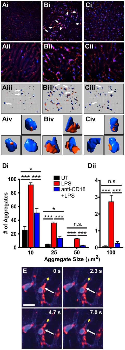
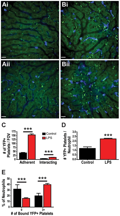
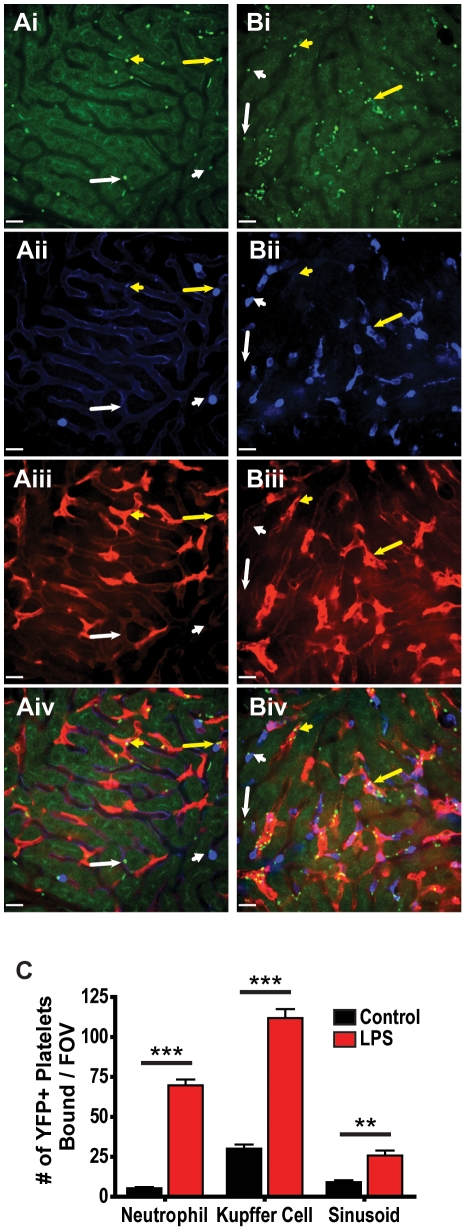
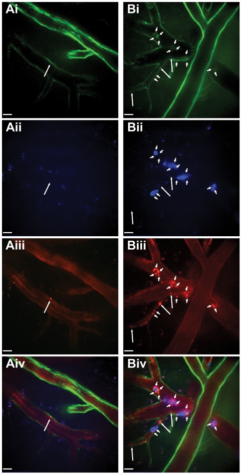
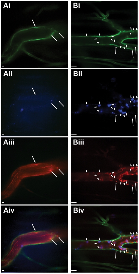
Platelets are central players in inflammation and are an important component of the innate immune response. The ability to visualize platelets within the live host is essential to understanding their role in these processes. Past approaches have involved adoptive transfer of labelled platelets, non-specific dyes, or the use of fluorescent antibodies to tag platelets in vivo. Often, these techniques result in either the activation of the platelet, or blockade of specific platelet receptors. In this report, we describe two new methods for intravital visualization of platelet biology, intravenous administration of labelled anti-CD49b, which labels all platelets, and CD41-YFP transgenic mice, in which a percentage of platelets express YFP. Both approaches label endogenous platelets and allow for their visualization using spinning-disk confocal fluorescent microscopy. Following LPS-induced inflammation, we were able to measure a significant increase in both the number and size of platelet aggregates observed within the vasculature of a number of different tissues. Real-time observation of these platelet aggregates reveals them to be large, dynamic structures that are continually expanding and sloughing-off into circulation. Using these techniques, we describe for the first time, platelet recruitment to, and behaviour within numerous tissues of the mouse, both under control conditions and following LPS induced inflammation.


Conflict of interest statement

Competing Interests: The authors have declared that no competing interests exist.

Figures

Figure 1
Figure 1. In vivo labelling of mouse platelets by intravenous injection of the anti-CD49b antibody HMα2.
Anti-CD49b uniformly labels all CD41+ platelets in mouse blood (A). Intravenous injection of PE-conjugated anti-CD49b (red) labels circulating platelets within the mouse liver (Bi –10× objective, Bii –20× objective) and pre-treatment of mice with anti-thrombocyte serum (Ci –10× objective, Cii –20× objective) results in the loss of CD49b+ particles confirming particles labelled by CD49b are indeed platelets. Neutrophils labelled with Alexa Fluor 647-conjugated anti-Gr-1 (blue). All scale bars, 20 µm.
Figure 2
Figure 2. Quantification of platelet aggregation in the livers of mice treated with intravenous LPS using PE-conjugated anti-CD49b.
Intravital microscopy of livers from untreated mice (Ai–Aiv), LPS treated mice (Bi–Biv) and mice pre-treated with an anti-CD18 blocking mAb followed by LPS (Ci–Civ). Representative fields of view at low power (10× objective) magnification (Ai, Bi, Ci) illustrating platelet aggregation in response to LPS and inhibition of aggregation following pre-treatment with an anti-CD18 blocking Ab. Platelets labelled with PE-conjugated anti-CD49b (red); neutrophils labelled with Alexa Fluor 647-conjugated anti-Gr-1 (blue). Arrows denote large platelet aggregates. Higher power (20× objective) extended focus images (Aii, Bii, Cii) and 3D opacity models (Aiii, Biii, Ciii) rendered from 29–30 z planes illustrating the extent of platelet aggregation in LPS treated mice. All scale bars, 20 µm; grid 25.7 µm. Select models of neutrophil-platelet aggregates (Aiv, Biv, Civ) extracted (as indicated by the white arrows) and enlarged from panel (iii). (Di, Dii) Quantification of the number of platelet aggregates equal to, or larger than the indicated sizes. *** p<0.001, * p<0.05, n.s. not significant (E) Time lapse images of neutrophil-platelet interactions illustrating the dynamic nature of the platelet aggregates within the liver. Long white arrow identifies a large platelet aggregate tearing off of an adherent neutrophil, short yellow arrow indicates direction of blood flow within the sinusoid. Sequential images represent 2.3 s intervals. Scale bar, 20 µm.
Figure 3
Figure 3. Quantification of platelet recruitment to livers of mice treated with intravenous LPS using CD41-YFPki/+ mice.
Representative fields of view of livers from untreated (Ai, Aii) and LPS treated (Bi, Bii) CD41-YFPki/+ mice. Neutrophils are labelled with Alexa Fluor 647-conjugated anti-Gr-1 (blue); YFP+ platelets and liver autofluorescence (green). All scale bars, 20 µm. (C) Quantification of the number of adherent (stationary for ≥30 s) and interacting (present in the field of view for but not stationary for ≥30 s) YFP+ platelets per field of view of liver in untreated and LPS treated CD41-YFPki/+ mice. (D) Average number of YFP+ platelets adherent to each neutrophil within the livers of untreated and LPS treated CD41-YFPki/+ mice. (E) Percentage of neutrophils with zero or with three or more adherent platelets within the livers of untreated and LPS treated CD41-YFPki/+ mice. *** p<0.001.
Figure 4
Figure 4. Characterization of platelet binding in the livers of mice treated with intravenous LPS using CD41-YFPki/+ mice.
Representative fields of view of livers from untreated (A) and LPS (B) treated CD41-YFPki/+ mice. Visualization of YFP+ platelets and liver autofluorescence (Ai, Bi), neutrophils labelled with Alexa Fluor 647-conjugated anti-Gr-1 (Aii, Bii), Kupffer cells labelled with PE-conjugated anti-F4/80 (Aiii, Biii), and overlay of fluorescent channels (Aiv, Biv). Examples of platelets interacting with the liver sinusoid away from neutrophils or Kupffer cells (long white arrows), platelets interacting with neutrophils (short white arrows), platelets interacting with Kupffer cells (short yellow arrows), and platelets appearing to interact with both Kupffer cells and neutrophils (long yellow arrows). All scale bars, 20 µm. (C) Quantification of the number of YFP+ platelets bound to neutrophils, Kupffer cells and liver sinusoids per field of view within livers of untreated and LPS treated CD41-YFPki/+ mice. *** p<0.001, ** p<0.01.
Figure 5
Figure 5. Intravital visualization of platelets labelled with PE-conjugated anti-CD49b within the mouse brain.
Spinning-disk confocal-microscopic imaging of brain pial arterioles and postcapillary venules in untreated (A) and LPS treated mice (B). Visualization of endothelium with Alexa Fluor 488-conjugated anti-CD31 (Ai, Bi), neutrophils labelled with Alexa Fluor 647-conjugated anti-Gr-1 (Aii, Bii), platelets stained with PE-conjugated anti-CD49b (Aiii, Biii), and multi-channel overlay (Aiv, Biv). There was a marked absence of platelet or neutrophil adhesion in brain pial vessels of untreated mice (A), however, profound neutrophil and platelet recruitment within brain venules (faint CD31 staining), but not in brain pial arterioles (bright CD31 staining), following LPS treatment (B). Small arrows identify platelets interacting with neutrophils; large arrows identify platelets interacting with the endothelium. All scale bars, 20 µm.
Figure 6
Figure 6. Intravital visualization of platelets labelled with PE-conjugated anti-CD49b within the mouse cremaster muscle.
Spinning-disk confocal-microscopic imaging of cremaster postcapillary venules in untreated (A) and LPS treated mice (B). Visualization of endothelium with Alexa Fluor 488-conjugated anti-CD31 (Ai, Bi), neutrophils labelled with Alexa Fluor 647-conjugated anti-Gr-1 (Aii, Bii), platelets stained with PE-conjugated anti-CD49b (Aiii, Biii), and multi-channel overlay (Aiv, Biv). A small number of platelets are seen to interact with the postcapillary endothelium in the cremaster muscle under basal conditions (A). Neutrophil recruitment and platelet interactions increase dramatically in the vessels of the cremaster muscle following LPS treatment (B). Small arrows identify platelets interacting with neutrophils; large arrows identify platelets interacting with the endothelium. All scale bars, 20 µm.

References

    1. Semple JW, Freedman J. Platelets and innate immunity. Cell Mol Life Sci. 2010;67:499–511. - PMC - PubMed
    1. Andonegui G, Kerfoot SM, McNagny K, Ebbert KV, Patel KD, et al. Platelets express functional Toll-like receptor-4. Blood. 2005;106:2417–2423. - PubMed
    1. Iannacone M, Sitia G, Isogawa M, Marchese P, Castro MG, et al. Platelets mediate cytotoxic T lymphocyte-induced liver damage. Nat Med. 2005;11:1167–1169. - PMC - PubMed
    1. Qu Z, Chaikof EL. Interface between hemostasis and adaptive immunity. Curr Opin Immunol. 2010;22:634–642. - PMC - PubMed
    1. Von Hundelshausen P, Koenen RR, Weber C. Platelet-mediated enhancement of leukocyte adhesion. Microcirculation. 2009;16:84–96. - PubMed

Publication types

MeSH terms