. 2012 Apr;165(7):2178-90.
doi: 10.1111/j.1476-5381.2011.01681.x.

Sulphide quinone reductase contributes to hydrogen sulphide metabolism in murine peripheral tissues but not in the CNS

Affiliations

Sulphide quinone reductase contributes to hydrogen sulphide metabolism in murine peripheral tissues but not in the CNS

D R Linden et al. Br J Pharmacol. 2012 Apr.

Abstract

Background and purpose: Hydrogen sulphide (H(2) S) is gaining acceptance as a gaseous signal molecule. However, mechanisms regarding signal termination are not understood. We used stigmatellin and antimycin A, inhibitors of sulphide quinone reductase (SQR), to test the hypothesis that the catabolism of H(2) S involves SQR.

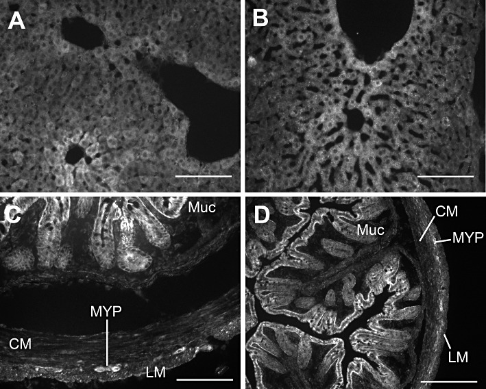
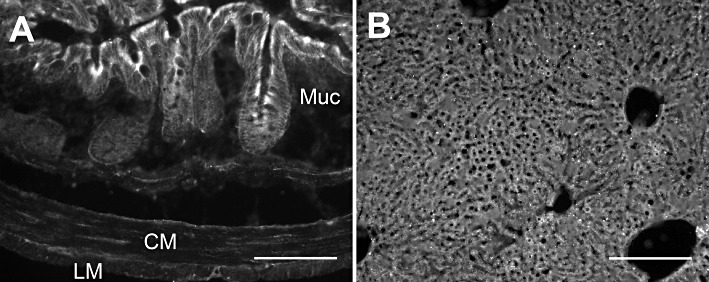
Experimental approach: H(2) S production and consumption were determined in living and intact mouse brain, liver and colonic muscularis externa using gas chromatography and HPLC. Expressions of SQR, ethylmalonic encephalopathy 1 (Ethe1) and thiosulphate transferase (TST; rhodanese) were determined by RT-PCR and immunohistochemistry.

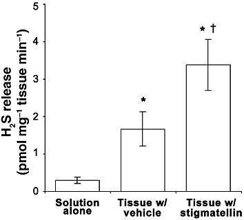
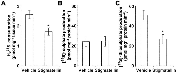
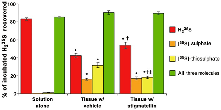
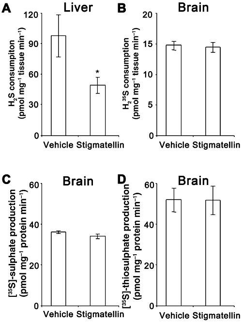
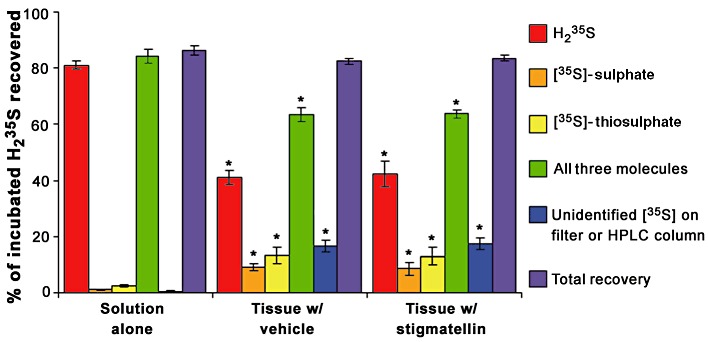
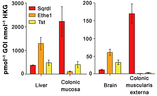
Key results: In the colonic muscularis externa, H(2) (35) S was catabolized to [(35) S]-thiosulphate and [(35) S]-sulphate, and stigmatellin reduced both the consumption of H(2) (35) S and formation of [(35) S]-thiosulphate. Stigmatellin also enhanced H(2) S release by the colonic muscularis externa. In the brain, catabolism of H(2) (35) S to [(35) S]-thiosulphate and [(35) S]-sulphate, which was stigmatellin-insensitive, partially accounted for H(2) (35) S consumption, while the remainder was captured as unidentified (35) S that was probably bound to proteins. Levels of mRNA encoding SQR were higher in the colonic muscularis externa and the liver than in the brain.

Conclusions and implications: These data support the concept that termination of endogenous H(2) S signalling in the colonic muscularis externa occurs via catabolism to thiosulphate and sulphate partially via a mechanism involving SQR. In the brain, it appears that H(2) S signal termination occurs partially through protein sequestration and partially through catabolism not involving SQR. As H(2) S has beneficial effects in animal models of human disease, we suggest that selective inhibition of SQR is an attractive target for pharmaceutical development.


Figures

Figure 1
Figure 1
The rate of H2S generation in normal Krebs solution containing 10 mM cysteine (solution alone), and the rate of H2S release in the same solution containing colonic muscularis externa with vehicle (0.1% EtOH) alone or with 3 µM stigmatellin. H2S release was greater in the presence of tissue, which was further increased by the presence of stigmatellin. Data are the mean ± SEM values for three independent experiments run in triplicate. *P < 0.05 compared with no tissue control; †P < 0.05 compared with vehicle treated control; repeated-measures anova, Neuman–Keuls post test.
Figure 2
Figure 2
The rates of consumption of H235S (A) and conversion of H235S to [35S]-sulphate (B) and [35S]-thiosulphate (C) by colonic muscularis externa incubated with vehicle (0.1% EtOH) alone or with 3 µM stigmatellin. Stigmatellin reduced H235S consumption and the conversion of H235S to [35S]-thiosulphate production but did not affect the conversion of H235S to [35S]-sulphate. Data are the mean ± SEM values for six independent experiments run in duplicate. *P < 0.05 compared with vehicle-treated control; paired t-test.
Figure 3
Figure 3
The proportion of H235S delivered to colonic muscularis externa incubated with vehicle (0.1% EtOH) or 3 µM stigmatellin that was recovered as either [35S]-sulphate, [35S]-thiosulphate or H235S or all three of these molecules. Nearly all of the supplied H235S was recovered as H235S when added to syringes containing the incubation solution without tissue (solution alone). The presence of tissue reduced the proportion of supplied H235S recovered as H235S and increased the proportion of H235S recovered as [35S]-sulphate and [35S]-thiosulphate (*P < 0.05 compared with solution alone, Kruskal–Wallis test followed by Dunn's test). Stigmatellin (3 µM) increased the proportion of H235S recovered as H235S (†P < 0.05 compared with tissue with vehicle, Kruskal–Wallis test followed by Dunn's test) and decreased the proportion of H235S recovered as [35S]-thiosulphate (‡P < 0.05 compared with tissue with vehicle, Mann–Whitney U-test) but did not affect the proportion of H235S recovered as [35S]-sulphate (P > 0.05 compared with tissue with vehicle, Kruskal–Wallis test and Mann–Whitney U-test). Data are the mean ± SEM proportions for 8–12 observations.
Figure 4
Figure 4
The rate of H2S consumption in liver tissue (A) and H235S consumption in brain tissue (B) incubated with vehicle (0.1% EtOH) alone or with 3 µM stigmatellin. Stigmatellin reduced H2S consumption by the liver but did not alter H235S consumption by brain tissue. Brain tissue was further analysed for the conversion of H235S to [35S]-sulphate (C) and [35S]-thiosulphate (D). Stigmatellin had no significant effect on the conversion of H235S to [35S]-sulphate or [35S]-thiosulphate. Data are the mean ± SEM values for three (liver) or five (brain) independent experiments run in duplicate. *P < 0.05 compared with vehicle-treated control; paired t-test.
Figure 5
Figure 5
The proportion of H235S delivered to brain tissue with vehicle (0.1% EtOH) or 3 µM stigmatellin that was recovered as either [35S]-sulphate, [35S]-thiosulphate or H235S, all three of these molecules, unidentified 35S recovered from the filter (>30 kDa) or HPLC column or a combination of the three identified molecules, and the 35S that was not identified. Nearly all of the supplied H235S was recovered as H235S when added to syringes containing the incubation solution without tissue (solution alone). Brain tissue reduced the proportion of H235S recovered as H235S and increased the proportions of H235S recovered as [35S]-sulphate, [35S]-thiosulphate, and unidentified [35S] (*P < 0.05 compared with solution alone, Kruskal–Wallis test followed by Dunn's test). Stigmatellin (3 µM) had no effect on any of these measurements. Data are the mean ± SEM proportions for 4–10 observations.
Figure 6
Figure 6
Expression of mRNA encoding SQR in the mouse liver, colonic mucosa and colonic muscularis externa, mRNA encoding Ethe1 in the mouse liver, colonic mucosa and brain, and mRNA encoding Tst in the mouse liver and brain. SQR, Ethe1 and Tst were expressed at low levels in brain tissue. Likewise, Ethe1 and Tst mRNA levels were at the detection limit in the colonic muscularis externa. Data are the mean ± SEM molar content of RNA encoding each gene of interest (GOI) in pmol, normalized to the average molar expression of RNA encoding β-actin and GAPDH as HKG in nmol for n= 3–4 animals.
Figure 7
Figure 7
Representative micrographs illustrating immunoreactivity for SQR in the mouse liver (A and B) and colon (C and D). In the liver, both the Sigma antiserum (A) and Proteintech antiserum (B) produced immunoreactivity in hepatocytes. Likewise staining for both antisera were similar in the colon with strong immunoreactivity in epithelial cells and cells within the muscle layers and myenteric plexus (C and D) (Sigma: C; Proteintech: D). (Muc: colonic mucosa; CM: circular muscle layer of the colon; LM: longitudinal muscle layer of the colon; MYP: myenteric plexus of the colon.) Scale bars illustrate 100 µm.
Figure 8
Figure 8
Representative micrographs illustrating immunoreactivity for Ethe1 in the mouse colon (A) and liver (B). Immunoreactivity was strong in the epithelial cells of the colonic mucosa while weak in the colonic muscularis externa. In the liver, there was strong immunoreactivity in hepatocytes. Scale bars illustrate 100 µm.

References

    1. Abe K, Kimura H. The possible role of hydrogen sulfide as an endogenous neuromodulator. J Neurosci. 1996;16:1066–1071. - PMC - PubMed
    1. Alexander SPH, Mathie A, Peters JA. Guide to Receptors and Channels (GRAC), 5th Edition. Br J Pharmacol. 2011;164(Suppl. 1):S1–S324. - PMC - PubMed
    1. Armstrong JS, Yang H, Duan W, Whiteman M. Cytochrome bc(1) regulates the mitochondrial permeability transition by two distinct pathways. J Biol Chem. 2004;279:50420–50428. - PubMed
    1. Bartholomew TC, Powell GM, Dodgson KS, Curtis CG. Oxidation of sodium sulphide by rat liver, lungs and kidney. Biochem Pharmacol. 1980;29:2431–2437. - PubMed
    1. Belyaeva EA, Dymkowska D, Wieckowski MR, Wojtczak L. Reactive oxygen species produced by the mitochondrial respiratory chain are involved in Cd2+-induced injury of rat ascites hepatoma AS-30D cells. Biochim Biophys Acta. 2006;1757:1568–1574. - PubMed

Publication types

MeSH terms