

doi: 10.1111/j.1749-6632.2011.06073.x.
The esophagogastric junction
Affiliations
- PMID: 21950822
- PMCID: PMC3276069
- DOI: 10.1111/j.1749-6632.2011.06073.x
Item in Clipboard
The esophagogastric junction
Ann N Y Acad Sci.
2011 Sep.
Abstract
The following discussion of the esophagogastric junctions includes commentaries on the three component structures of the sphincteric segment between the stomach and the esophagus; the pressure contributions from the three sphincteric components in normal subjects and in gastroesophageal reflux (GERD) patients; the mechanism of action of endoscopic plication to determine the underlying pathophysiology of GERD; and in vitro muscle strip studies of defects within the gastroesophageal sphincteric segment potentially leading to GERD.
© 2011 New York Academy of Sciences.
Conflict of interest statement
The authors declare no conflicts of interest.
Figures
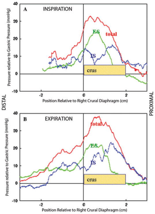
Panels A and B show average pressure curves from the same group of 15 normal subjects, referenced to distal margin of the right crus muscles Panel A is for full inspiration and Panel B for full expiration. The red curves are averaged pressure before administering atropine, the green curves are post atropine pressure and the blue curve is the average of the difference between pre and post atropine pressures (effectively the red curve minus the green curve). The vertical line at zero is the lower margin of the right crural diaphragm.
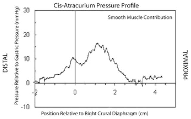
Average pressure curve in full inspiration, referenced to the lower margin of the right crus muscles, from seven subjects undergoing surgery for nongastric or esophageal pathology after giving Cis-atracurium to paralyze the skeletal muscle.
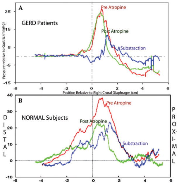
Similarly to Figure 1, Panel A shows average pressure curves in full expiration from seven GERD patients, referenced to right crural diaphragm. Panel B shows the average pressure curves in full expiration from 15 normal control subjects from Figure 1. The red curve shows average pressure preatropine pressure and the green curve post atropine. The blue curve is the subtraction curve (red curve minus green curve). The vertical line at zero is the lower margin of the right crus muscles.
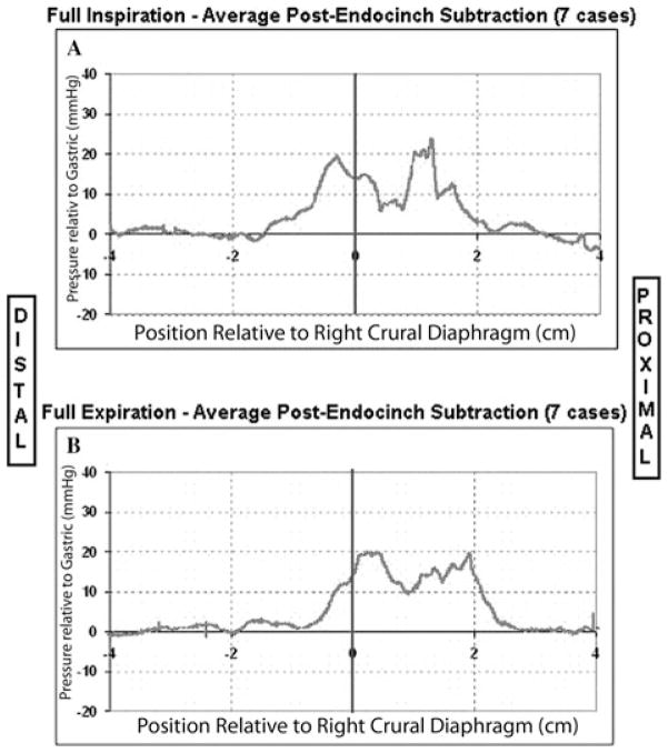
The average subtraction pressure curves from seven GERD patients after the Endocinch procedure in full inspiration (Panel A) and full expiration (Panel B) procedure.
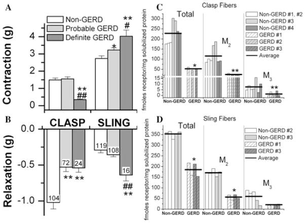
(A) and (B) show that clasp fibers from two definite GERD donors contracted less in response to maximally effective carbachol concentrations than 11 non-GERD and four probable GERD donors, whereas clasp fibers from both probable and definite GERD donors relaxed less than non-GERD donors. Sling fibers of both probable and definite GERD donors contracted greater than non-GERD donors, and sling fibers from definite GERD donors relaxed more than non-GERD donors. The density of M2 and M3 muscarinic receptors, determined by immunoprecipitation, was lower in clasp fibers of GERD donors (C) and M2 density was lower in sling fibers of GERD donors (D).
References
-
- Ingelfinger FJ. Esophageal motility. Physiol Rev. 1958;38:533–584. - PubMed
-
- Boyle JT, Altschuler SM, Nixon TE, et al. Role of the diaphragm in the genesis of the lower esophageal sphincter pressure in the cat. Gastroenterology. 1985;88:723–730. - PubMed
-
- Code CF, Fyke FE, Jr, Schlegel JF, et al. The gastroesophageal sphincter in healthy human beings. Gastroenterologia. 1956;86:135–150. - PubMed
-
- Liebermann-Meffert D, AllgÖwer M, Schmid P, Blum AL. Muscular equivalent of the lower esophageal sphincter. Gastroenterology. 1979;76:31–38. - PubMed
MeSH terms
Grants and funding
LinkOut - more resources
Full Text Sources
