. 2011 Nov 8;50(44):9664-80.
doi: 10.1021/bi201368u. Epub 2011 Oct 13.

Functional consequences of the creation of an Asp-His-Fe triad in a 3/3 globin

Affiliations

Functional consequences of the creation of an Asp-His-Fe triad in a 3/3 globin

Edward L D'Antonio et al. Biochemistry. .

Abstract

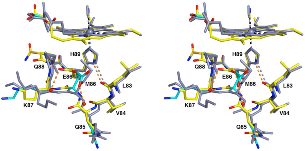
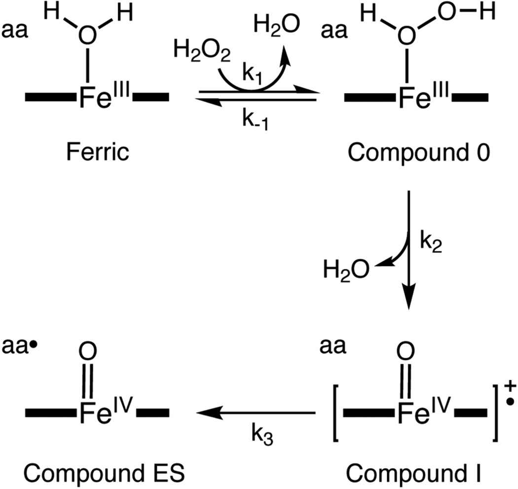
The proximal side of dehaloperoxidase-hemoglobin A (DHP A) from Amphitrite ornata has been modified via site-directed mutagenesis of methionine 86 into aspartate (M86D) to introduce an Asp-His-Fe triad charge relay. X-ray crystallographic structure determination of the metcyano forms of M86D [Protein Data Bank (PDB) entry 3MYN ] and M86E (PDB entry 3MYM ) mutants reveal the structural origins of a stable catalytic triad in DHP A. A decrease in the rate of H(2)O(2) activation as well as a lowered reduction potential versus that of the wild-type enzyme was observed in M86D. One possible explanation for the significantly lower activity is an increased affinity for the distal histidine in binding to the heme Fe to form a bis-histidine adduct. Resonance Raman spectroscopy demonstrates a pH-dependent ligation by the distal histidine in M86D, which is indicative of an increased trans effect. At pH 5.0, the heme Fe is five-coordinate, and this structure resembles the wild-type DHP A resting state. However, at pH 7.0, the distal histidine appears to form a six-coordinate ferric bis-histidine (hemichrome) adduct. These observations can be explained by the effect of the increased positive charge on the heme Fe on the formation of a six-coordinate low-spin adduct, which inhibits the ligation and activation of H(2)O(2) as required for peroxidase activity. The results suggest that the proximal charge relay in peroxidases regulate the redox potential of the heme Fe but that the trans effect is a carefully balanced property that can both activate H(2)O(2) and attract ligation by the distal histidine. To understand the balance of forces that modulate peroxidase reactivity, we studied three M86 mutants, M86A, M86D, and M86E, by spectroelectrochemistry and nuclear magnetic resonance spectroscopy of (13)C- and (15)N-labeled cyanide adducts as probes of the redox potential and of the trans effect in the heme Fe, both of which can be correlated with the proximity of negative charge to the N(δ) hydrogen of the proximal histidine, consistent with an Asp-His-Fe charge relay observed in heme peroxidases.


Figures

Figure 1
Figure 1
Structural comparison of the Asp-His-Fe triads observed in DHP A(M86D) and wild-type CcP. Superposition of chain B for M86D (PDB 3MYN) (coloring scheme: C = yellow, O = red, N = blue, Fe3+ = blue sphere, hydrogen bond = green dashed line; carbon atoms are either colored gold or cyan for equal or lower occupancy conformers, respectively) and wild-type CcP (PDB 1CCA) (14) (all atoms and interactions are colored grey). The D86-H89 hydrogen bond in M86D has an O---N separation of 2.9 Å and the D235-H175 hydrogen bond in CcP has an O---N separation of 3.0 Å. The superposition is based on the alignment of the heme Fe and the proximal histidine Nε atom between the two structures.
Figure 2
Figure 2
DHP-M86D-CN complex, pH 6.5. (A) Simulated annealing omit maps (blue) are contoured at 2.5σ for residues D86 and H89. H89 is found rotated substantially from the conformation in the wild type structure and forms a hydrogen bond with D86 (O---N separation is 2.9 Å). Atoms are represented by the following coloring scheme: C = yellow, O = red, N = blue, Fe3+ = marine blue sphere, solvent = red spheres; carbon atoms are colored as cyan and gold in residue side chains with lower and equal occupancy conformers, respectively. Iron coordination interactions are indicated by black dashed lines and hydrogen bonds are indicated as orange dashed lines. (B) Superposition of chain B for M86D-CN (same coloring scheme as in (A)) and chain A for DHP A-CN (X-ray data from a rotating anode source (unpublished results), all atoms colored grey).
Figure 3
Figure 3
DHP-M86E-CN complex, pH 6.5. Superposition of chain B for M86E-CN (coloring scheme: C = yellow, O = red, N = blue, Fe3+ = marine blue sphere, solvent = red spheres, iron coordination = black dashed lines, hydrogen bonds = orange dashed lines; carbon atoms are colored as cyan and gold in residue side chains with lower and equal occupancy conformers, respectively) and chain A for DHP A-CN (X-ray data from a rotating anode source (unpublished results), all atoms colored grey). E86 is solvent exposed and does not interact with H89 and H89 remains hydrogen bonded with the backbone carbonyl of L83.
Figure 4
Figure 4
UV-VIS thin-layer spectroelectrochemistry of M86D. (A) Spectra are shown for M86D in 100 mM potassium phosphate (pH 7.0), 25 °C, [Ru(NH3)6]3+ mediator, at applied potentials (EAPP vs. SHE): (a) −0.303 V, (b) −0.013 V, (c) +0.007 V, (d) +0.027 V, (e) +0.047 V, (f) +0.067 V, (g) +0.087 V, (h) +0.117 V and (i) +0.197 V. (B) Nernst plot of the same data.
Figure 5
Figure 5
Structural comparison of the DHP A proximal regions mutants (A) M86E-CN and (B) M86D-CN. Relevant hydrogen bonding distances are shown. For clarity, only the shortest distance between (D86)Oδ---(H89)Nδ is shown in panel (B) and the heme Fe ion cyanide ligand was omitted in both panels.
Figure 6
Figure 6
13C paramagnetic NMR spectra of various heme proteins. Spectra are shown for 13CN bound to the heme Fe of (A) heme proteins: (a) DHP A, (b) HHMb, (c) HRP-type I, and (B) comparison of (a) DHP A with the M86 mutants, (b) M86A, (c) M86E, and (d) M86D.
Figure 7
Figure 7
Single-mixing stopped-flow kinetics of the DHP A(M86E) mutant with hydrogen peroxide. (A) Single-mixing stopped-flow UV-VIS spectroscopic monitoring of the reaction (900 scans, 85 sec) between DHP A(M86E) (10 µM) and a 10-fold excess of H2O2 at pH 7.0, see experimental for details. (B) Calculated UV-VIS spectra for resting (black), Compound ES (red), and the putative species Compound RH (blue) of DHP A(M86E) are shown; the rapid-scanning data from panel (A) were compiled and fitted to a double exponential reaction model using the Specfit global analysis program. (C) Relative concentration profile determined from the three component fit used in panel (B).
Figure 8
Figure 8
Resonance Raman spectra of M86D in the presence and absence of cyanide ion. (A) The ferric forms of (a) M86D-CN and (b) M86D are shown at pH 7.0. (B) The ferric forms of M86D are shown at (a) pH 7.0, (b) pH 6.0, and (c) pH 5.0.
Figure 9
Figure 9
Potential energy surfaces along the Fe-Nε bond for 4-methyl imidazole binding to the heme Fe. (A) Bis-(4-methyl imidazole) adducts for various charged heme species. (B) Plot of Fe-Nε binding energy as a function of the Fe---Nε bond distance. Coloring scheme for the adducts: ferrous neutral = red, ferric neutral = violet, ferric anion (Imdistal) = blue, ferric anion (Improximal) = black, and the ferric dianion = brown.
Figure 10
Figure 10
Molecular dynamics simulation of the M86D mutant Fe---Nδ (red) and Fe---Nε (blue) with a charge set for the heme that accounts for ferric heme.
Scheme I
Scheme I
The 2-electron oxidation of ferric DHP A to Compound ES that is initiated by hydrogen peroxide binding to the heme Fe.

References

    1. Chen YP, Woodin SA, Lincoln DE, Lovell CR. An unusual dehalogenating peroxidase from the marine terebellid polychaete amphitrite ornata. J. Biol. Chem. 1996;271:4609–4612. - PubMed
    1. Lebioda L, LaCount MW, Zhang E, Chen YP, Han KP, Whitton MM, Lincoln DE, Woodin SA. An enzymatic globin from a marine worm. Nature. 1999;401:445. - PubMed
    1. LaCount MW, Zhang EL, Chen YP, Han KP, Whitton MM, Lincoln DE, Woodin SA, Lebioda L. The crystal structure and amino acid sequence of dehaloperoxidase from amphitrite ornata indicate common ancestry with globins. J. Biol. Chem. 2000;275:18712–18716. - PubMed
    1. Belyea J, Gilvey LB, Davis MF, Godek M, Sit TL, Lommel SA, Franzen S. Enzyme function of the globin dehaloperoxidase from amphitrite ornata is activated by substrate binding. Biochemistry. 2005;44:15637–15644. - PubMed
    1. Bashford D, Chothia C, Lesk AM. Determinants of a protein fold - unique features of the globin amino-acid-sequences. J. Mol. Biol. 1987;196:199–216. - PubMed

Publication types