

Optical scattering coefficient estimated by optical coherence tomography correlates with collagen content in ovarian tissue
- PMID: 21950907
- PMCID: PMC3194791
- DOI: 10.1117/1.3625247
Optical scattering coefficient estimated by optical coherence tomography correlates with collagen content in ovarian tissue
Abstract
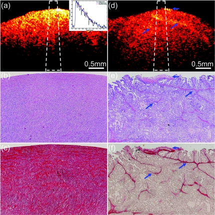
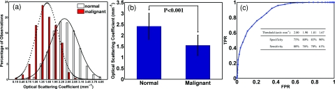
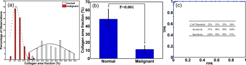
Optical scattering coefficient from ex vivo unfixed normal and malignant ovarian tissue was quantitatively extracted by fitting optical coherence tomography (OCT) A-line signals to a single scattering model. 1097 average A-line measurements at a wavelength of 1310 nm were performed at 108 sites obtained from 18 ovaries. The average scattering coefficient obtained from the normal tissue group consisted of 833 measurements from 88 sites was 2.41 mm(-1) (± 0.59), while the average coefficient obtained from the malignant tissue group consisted of 264 measurements from 20 sites was 1.55 mm(-1) (± 0.46). The malignant ovarian tissue showed significant lower scattering than the normal group (p < 0.001). The amount of collagen within OCT imaging depth was analyzed from the tissue histological section stained with Sirius Red. The average collagen area fraction (CAF) obtained from the normal tissue group was 48.4% (± 12.3%), while the average CAF obtained from the malignant tissue group was 11.4% (± 4.7%). A statistical significance of the collagen content was found between the two groups (p < 0.001). These results demonstrated that quantitative measurements of optical scattering coefficient from OCT images could be a potential powerful method for ovarian cancer detection.
Figures
Similar articles
-
Quantitative analysis of angle-resolved scattering properties of ovarian tissue using optical coherence tomography.J Biomed Opt. 2012 Sep;17(9):90503-1. doi: 10.1117/1.JBO.17.9.090503. J Biomed Opt. 2012. PMID: 23085900 Free PMC article.
-
Quantitative analysis of estimated scattering coefficient and phase retardation for ovarian tissue characterization.Biomed Opt Express. 2012 Jul 1;3(7):1548-56. doi: 10.1364/BOE.3.001548. Epub 2012 Jun 7. Biomed Opt Express. 2012. PMID: 22808427 Free PMC article.
-
Characterizing optical properties and spatial heterogeneity of human ovarian tissue using spatial frequency domain imaging.J Biomed Opt. 2016 Oct;21(10):101402. doi: 10.1117/1.JBO.21.10.101402. J Biomed Opt. 2016. PMID: 26822943 Free PMC article.
-
An overview of optical coherence tomography for ovarian tissue imaging and characterization.Wiley Interdiscip Rev Nanomed Nanobiotechnol. 2015 Jan-Feb;7(1):1-16. doi: 10.1002/wnan.1306. Epub 2014 Oct 20. Wiley Interdiscip Rev Nanomed Nanobiotechnol. 2015. PMID: 25329515 Free PMC article. Review.
-
Review of methods and applications of attenuation coefficient measurements with optical coherence tomography.J Biomed Opt. 2019 Sep;24(9):1-17. doi: 10.1117/1.JBO.24.9.090901. J Biomed Opt. 2019. PMID: 31520468 Free PMC article. Review.
Cited by
-
Optical coherence tomography can assess skeletal muscle tissue from mouse models of muscular dystrophy by parametric imaging of the attenuation coefficient.Biomed Opt Express. 2014 Mar 19;5(4):1217-32. doi: 10.1364/BOE.5.001217. eCollection 2014 Apr 1. Biomed Opt Express. 2014. PMID: 24761302 Free PMC article.
-
Multimodal evaluation of ultra-short laser pulses treatment for skin burn injuries.Biomed Opt Express. 2017 Feb 16;8(3):1575-1588. doi: 10.1364/BOE.8.001575. eCollection 2017 Mar 1. Biomed Opt Express. 2017. PMID: 28663850 Free PMC article.
-
Accurate attenuation characterization in optical coherence tomography using multi-reference phantoms and deep learning.Biomed Opt Express. 2024 Nov 6;15(12):6697-6714. doi: 10.1364/BOE.543606. eCollection 2024 Dec 1. Biomed Opt Express. 2024. PMID: 39679392 Free PMC article.
-
Bayesian analysis of depth resolved OCT attenuation coefficients.Sci Rep. 2021 Jan 26;11(1):2263. doi: 10.1038/s41598-021-81713-7. Sci Rep. 2021. PMID: 33500435 Free PMC article.
-
Characterization of the Optical Properties of Photoluminescent Turbid Media Using an Integrating Sphere and Monte Carlo Simulations.Materials (Basel). 2024 Dec 12;17(24):6072. doi: 10.3390/ma17246072. Materials (Basel). 2024. PMID: 39769671 Free PMC article.
References
-
- Brewer M. A., et al., “Imaging of the ovary,” Technol. Cancer Res. Treat. 3, 617–627 (2004). - PubMed
Publication types
MeSH terms
Substances
Grants and funding
LinkOut - more resources
Full Text Sources
Medical
