. 2011 Sep:1233:91-9.
doi: 10.1111/j.1749-6632.2011.06168.x.

Sources of calretinin inputs to motoneurons of extraocular muscles involved in upgaze

Affiliations

Sources of calretinin inputs to motoneurons of extraocular muscles involved in upgaze

Julia Ahlfeld et al. Ann N Y Acad Sci. 2011 Sep.

Abstract

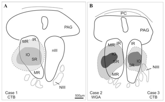
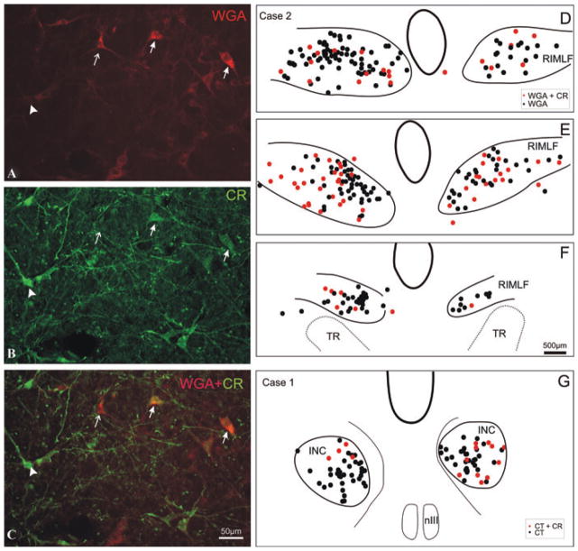
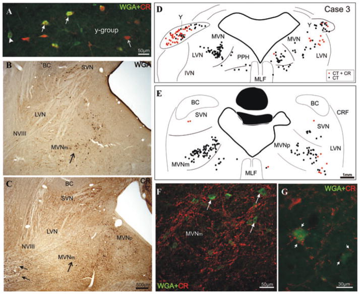
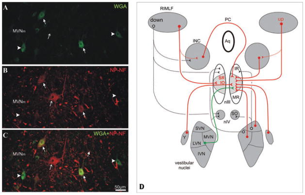
Recent monkey studies showed that motoneurons of the oculomotor nucleus involved in upward eye movements receive a selective input from afferents containing calretinin (CR). Here, we investigated the sources of these CR-positive afferents. After injections of tract-tracers into the oculomotor nucleus (nIII) of two monkeys, the retrograde labeling was combined with CR-immunofluorescence in frozen brainstem sections. Three sources of CR inputs to nIII were found: the rostral interstitial nucleus of the medial longitudinal fascicle (RIMLF), the interstitial nucleus of Cajal, and the y-group. CR is not present in all premotor upward-moving pathways. The excitatory secondary vestibulo-ocular neurons in the magnocellular part of the medial vestibular nuclei contained nonphosphorylated neurofilaments, but no CR, and they received a strong supply of large CR-positive boutons. In conclusion, the present study presents evidence that only specific premotor pathways for upward eye movements--excitatory upgaze pathways--contain CR, but not the up vestibulo-ocular reflex pathways. This property may help to differentiate between premotor up- and downgaze pathways in correlative clinico-anatomical studies in humans.


Conflict of interest statement

Conflicts of interest

The authors declare no conflicts of interest.

Figures

Figure 1
Figure 1
Schematic drawings of transverse sections through the monkey oculomotor nucleus (nIII) to show the location of tracer injection sites (dark) and the extent of the uptake area (light gray) in three cases. NIII, oculomotor nerve; PAG, periaqueductal gray; PC, posterior commissure.
Figure 2
Figure 2
High-power magnification of a transverse section through the rostral interstitial nucleus of the medial longitudinal fascicle (RIMLF) showing the tracer-labeled (WGA) premotor neurons (red; A) and calretinin (CR)-positive neurons (green; B). In the superposition of A and B, double-labeled neurons appear in orange (solid arrows), clearly distinguished from neurons containing either only CR (green, arrow head) or the tracer WGA (red, arrow; C). Plots of transverse sections through the RIMLF (D–F) and interstitial nucleus of Cajal (INC; G) of a monkey with a CTB-injection into the left oculomotor nucleus (nIII) in rostrocaudal order (D–F): tracer-labeled CR-positive putative upburst neurons (red dots) are intermingled with tracer-labeled CR-negative putative down-burst neurons (black dots). TR, tractus retroflexus.
Figure 3
Figure 3
Superposition of high-power photographs of a transverse section through the y-group showing premotor tracer-labeled neurons (green, arrowhead), some of which contain CR (orange, solid arrow). Not all CR-positive neurons are tracer labeled (red, thin arrow; A). Photographs of corresponding sections through the vestibular nuclei of monkey stained for retrograde tracer WGA (black cells; B) or CR (brown cells; C). Note that MVNm contains numerous tracer-labeled neurons (arrow; B), but few CR-positive neurons (arrow; C). The vestibular nerve (NVIII) contains numerous CR-positive fibers (solid arrows). Transverse sections through the vestibular nuclei of a monkey with a tracer (CTB) injection into the right oculomotor nucleus with the retrogradely labeled neurons plotted (black dots; D, E). Tracer-labeled CR-positive neurons (red dots) are mainly found in the y-group of both sides (D). Putative excitatory secondary vestibulo-ocular neurons in the left MVm lack CR (E), but they are contacted by numerous large CR-positive terminals (red, arrows; F, G).
Figure 4
Figure 4
High-power magnification of a transverse section through the magnocellular part of the medial vestibular nucleus (MVNm) showing the WGA-labeled secondary vestibulo-ocular neurons (A) and neurons stained for nonphosphorylated neurofilaments (NP-NF; B). The superposition reveals that many, but not all, vestibulo-ocular neurons (green, arrowhead) are NP-NF–positive (solid arrow) and not all NP-NF–positive neurons are tracer labeled (red, thin arrow; C). Simplified diagram summarizing the premotor pathways for vertical eye movements. The projections found after a tracer injection into the right oculomotor nucleus (nIII) and associated with CR found are drawn in red, and those associated with NP-NF are drawn in green (D). IVN, inferior vestibular nucleus; LVN, lateral vestibular nucleus; PC, posterior commissure.

References

    1. Schulze C, Mustari MJ, Holstein GR, Horn AKE. Transmitter inputs to different motoneuron subgroups in the oculomotor and trochlear nucleus in monkey. Soc Neurosci Abstr. 2009;35:356–359. - PMC - PubMed
    1. Leigh RJ, Zee DS. The Neurology of Eye Movements. 4. Oxford Univ. Press; New York: 2006.
    1. Büttner-Ennever JA, Gerrits NM. Vestibular System. In: Paxinos G, Mai JK, editors. The Human Nervous System. Elsevier Academic Press; Amsterdam: 2004. pp. 1212–1240.
    1. Highstein SM, Holstein GR. The anatomy of the vestibular nuclei. Prog Brain Res. 2006;151:157–203. - PubMed
    1. Horn AKE. The reticular formation. Prog Brain Res. 2006;151:127–155. - PubMed

Publication types

MeSH terms